2OD9 | pdb_00002od9

Structural Basis for Nicotinamide Inhibition and Base Exchange in Sir2 Enzymes


Experimental Data Snapshot

  • Method: X-RAY DIFFRACTION
  • Resolution: 2.05 Å
  • R-Value Free: 
    0.245 (Depositor), 0.226 (DCC) 
  • R-Value Work: 
    0.218 (Depositor), 0.227 (DCC) 
  • R-Value Observed: 
    0.245 (Depositor) 

Starting Model: experimental
View more details

wwPDB Validation 3D Report Full Report

Validation slider image for 2OD9

Ligand Structure Quality Assessment 


This is version 1.6 of the entry. See complete history

Literature

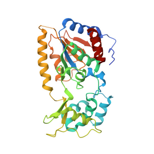
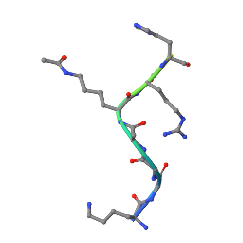
Structural basis for nicotinamide inhibition and base exchange in sir2 enzymes.

Sanders, B.D.Zhao, K.Slama, J.T.Marmorstein, R.

(2007) Mol Cell 25: 463-472

  • DOI: https://doi.org/10.1016/j.molcel.2006.12.022
  • Primary Citation Related Structures: 
    2OD2, 2OD7, 2OD9, 2QQF, 2QQG

  • PubMed Abstract: 

    The Sir2 family of proteins consists of broadly conserved NAD(+)-dependent deacetylases that are implicated in diverse biological processes, including DNA regulation, metabolism, and longevity. Sir2 proteins are regulated in part by the cellular concentrations of a noncompetitive inhibitor, nicotinamide, that reacts with a Sir2 reaction intermediate via a base-exchange reaction to reform NAD(+) at the expense of deacetylation. To gain a mechanistic understanding of nicotinamide inhibition in Sir2 enzymes, we captured the structure of nicotinamide bound to a Sir2 homolog, yeast Hst2, in complex with its acetyl-lysine 16 histone H4 substrate and a reaction intermediate analog, ADP-HPD. Together with related biochemical studies and structures, we identify a nicotinamide inhibition and base-exchange site that is distinct from the so-called "C pocket" binding site for the nicotinamide group of NAD(+). These results provide insights into the Sir2 mechanism of nicotinamide inhibition and have important implications for the development of Sir2-specific effectors.


  • Organizational Affiliation
    • The Wistar Institute, University of Pennsylvania, Philadelphia, PA 19104, USA.

Macromolecule Content 

  • Total Structure Weight: 37.14 kDa 
  • Atom Count: 2,527 
  • Modeled Residue Count: 298 
  • Deposited Residue Count: 322 
  • Unique protein chains: 2

Macromolecules

Find similar proteins by:|  3D Structure
Entity ID: 1
MoleculeChains  Sequence LengthOrganismDetailsImage
NAD-dependent deacetylase HST2308Saccharomyces cerevisiaeMutation(s): 0 
Gene Names: HST2
EC: 3.5.1 (PDB Primary Data), 2.3.1.286 (UniProt)
UniProt
Find proteins for P53686 (Saccharomyces cerevisiae (strain ATCC 204508 / S288c))
Explore P53686 
Go to UniProtKB:  P53686
Entity Groups
Sequence Clusters30% Identity50% Identity70% Identity90% Identity95% Identity100% Identity
UniProt GroupP53686
Sequence Annotations
Expand
Reference Sequence
Find similar proteins by:  Sequence   |   3D Structure  
Entity ID: 2
MoleculeChains  Sequence LengthOrganismDetailsImage
H4 peptide14N/AMutation(s): 0 

Small Molecules

Ligands 3 Unique
IDChains Name / Formula / InChI Key2D Diagram3D Interactions
A1R

Query on A1R


Download:Ideal Coordinates CCD File
D [auth A]5'-O-[(S)-{[(S)-{[(2R,3R,4S)-3,4-DIHYDROXYPYRROLIDIN-2-YL]METHOXY}(HYDROXY)PHOSPHORYL]OXY}(HYDROXY)PHOSPHORYL]ADENOSINE
C15 H24 N6 O12 P2
NDQDTBCXPOIQGT-UHNJQBFTSA-N
NCA

Query on NCA


Download:Ideal Coordinates CCD File
E [auth A]NICOTINAMIDE
C6 H6 N2 O
DFPAKSUCGFBDDF-UHFFFAOYSA-N
ZN

Query on ZN


Download:Ideal Coordinates CCD File
C [auth A]ZINC ION
Zn
PTFCDOFLOPIGGS-UHFFFAOYSA-N
Modified Residues  1 Unique
IDChains TypeFormula2D DiagramParent
ALY
Query on ALY
B
L-PEPTIDE LINKINGC8 H16 N2 O3LYS
Binding Affinity Annotations 
IDSourceBinding Affinity
NCA BindingDB:  2OD9 IC50: 9.10e+4 (nM) from 1 assay(s)

Experimental Data & Validation

Experimental Data

  • Method: X-RAY DIFFRACTION
  • Resolution: 2.05 Å
  • R-Value Free:  0.245 (Depositor), 0.226 (DCC) 
  • R-Value Work:  0.218 (Depositor), 0.227 (DCC) 
  • R-Value Observed: 0.245 (Depositor) 
Space Group: P 32 2 1
Unit Cell:
Length ( Å )Angle ( ˚ )
a = 105.94α = 90
b = 105.94β = 90
c = 67.1γ = 120
Software Package:
Software NamePurpose
DENZOdata reduction
SCALEPACKdata scaling
AMoREphasing
CNSrefinement
PDB_EXTRACTdata extraction
HKL-2000data collection
HKL-2000data reduction

Structure Validation

View Full Validation Report


Ligand Structure Quality Assessment 


Entry History 

Deposition Data

Revision History  (Full details and data files)

  • Version 1.0: 2007-02-27
    Type: Initial release
  • Version 1.1: 2008-05-01
    Changes: Version format compliance
  • Version 1.2: 2011-07-13
    Changes: Version format compliance
  • Version 1.3: 2017-10-18
    Changes: Refinement description
  • Version 1.4: 2023-08-30
    Changes: Data collection, Database references, Derived calculations, Refinement description
  • Version 1.5: 2023-11-15
    Changes: Data collection
  • Version 1.6: 2024-10-16
    Changes: Structure summary