NMR structural characterization of a minimal peptide antagonist bound to the extracellular domain of the corticotropin-releasing factor1 receptor.
Mesleh, M.F., Shirley, W.A., Heise, C.E., Ling, N., Maki, R.A., Laura, R.P.(2007) J Biological Chem 282: 6338-6346
- PubMed: 17192263 Search on PubMed
- DOI: https://doi.org/10.1074/jbc.M609816200
- Primary Citation Related Structures:
2O8Z - PubMed Abstract:
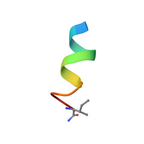
Natural peptide agonists of corticotrophin-releasing factor (CRF) receptors bind to the receptor by a two-site mechanism as follows: the carboxyl end of the ligand binds the N-terminal extracellular domain (ECD) of the receptor and the amino portion of the ligand binds the extracellular face of the seven transmembrane region. Recently, peptide antagonists homologous to the 12 C-terminal residues of CRF have been derived, which bind the CRF(1) receptor through an interaction with the ECD. Here we characterized the binding of a minimal 12-residue peptide antagonist while bound to the isolated ECD of the CRF(1) receptor. We have expressed and purified soluble and properly folded ECD independent from the seven-transmembrane region as a thioredoxin fusion protein in Escherichia coli. A model of the peptide antagonist, cyclic corticotrophin-releasing factor residues 30-41 (cCRF(30-41)), was calculated while bound to the recombinant ECD using transferred nuclear Overhauser effect spectroscopy. Although the peptide is unstructured in solution, it adopts an alpha-helical conformation when bound to the ECD. Residues of cCRF(30-41) comprising the binding interface with the ECD were mapped using saturation transfer difference NMR. Two hydrophobic residues (Met(38) and Ile(41)) as well as two amide groups (Asn(34) and the C-terminal amide) on one face of the helix defined the binding epitope of the antagonist. This epitope may be used as a starting point for development of non-peptide antagonists targeting the ECD of this receptor.
- Department of Medicinal Chemistry, Neurocrine Biosciences, Inc., San Diego, California 92130, USA.
Organizational Affiliation: