2O4I | pdb_00002o4i

Structure of TREX1 in complex with DNA


Experimental Data Snapshot

  • Method: X-RAY DIFFRACTION
  • Resolution: 3.50 Å
  • R-Value Free: 
    0.283 (Depositor), 0.275 (DCC) 
  • R-Value Work: 
    0.243 (Depositor), 0.237 (DCC) 
  • R-Value Observed: 
    0.245 (Depositor) 

Starting Model: experimental
View more details

wwPDB Validation 3D Report Full Report

Validation slider image for 2O4I

This is version 1.4 of the entry. See complete history

Literature

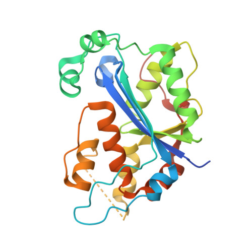
Structure of the dimeric exonuclease TREX1 in complex with DNA displays a proline-rich binding site for WW Domains.

Brucet, M.Querol-Audi, J.Serra, M.Ramirez-Espain, X.Bertlik, K.Ruiz, L.Lloberas, J.Macias, M.J.Fita, I.Celada, A.

(2007) J Biological Chem 282: 14547-14557

  • DOI: https://doi.org/10.1074/jbc.M700236200
  • Primary Citation Related Structures: 
    2O4G, 2O4I

  • PubMed Abstract: 

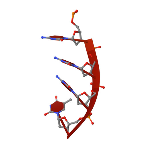
    TREX1 is the most abundant mammalian 3' --> 5' DNA exonuclease. It has been described to form part of the SET complex and is responsible for the Aicardi-Goutières syndrome in humans. Here we show that the exonuclease activity is correlated to the binding preferences toward certain DNA sequences. In particular, we have found three motifs that are selected, GAG, ACA, and CTGC. To elucidate how the discrimination occurs, we determined the crystal structures of two murine TREX1 complexes, with a nucleotide product of the exonuclease reaction, and with a single-stranded DNA substrate. Using confocal microscopy, we observed TREX1 both in nuclear and cytoplasmic subcellular compartments. Remarkably, the presence of TREX1 in the nucleus requires the loss of a C-terminal segment, which we named leucine-rich repeat 3. Furthermore, we detected the presence of a conserved proline-rich region on the surface of TREX1. This observation points to interactions with proline-binding domains. The potential interacting motif "PPPVPRPP" does not contain aromatic residues and thus resembles other sequences that select SH3 and/or Group 2 WW domains. By means of nuclear magnetic resonance titration experiments, we show that, indeed, a polyproline peptide derived from the murine TREX1 sequence interacted with the WW2 domain of the elongation transcription factor CA150. Co-immunoprecipitation studies confirmed this interaction with the full-length TREX1 protein, thereby suggesting that TREX1 participates in more functional complexes than previously thought.


  • Organizational Affiliation
    • Macrophage Biology Group, Institute for Research in Biomedicine, University of Barcelona, Barcelona, Spain.

Macromolecule Content 

  • Total Structure Weight: 69.29 kDa 
  • Atom Count: 3,517 
  • Modeled Residue Count: 444 
  • Deposited Residue Count: 544 
  • Unique protein chains: 1
  • Unique nucleic acid chains: 1

Macromolecules


Find similar proteins by:|  3D Structure
Entity ID: 2
MoleculeChains  Sequence LengthOrganismDetailsImage
Three prime repair exonuclease 1C [auth A],
D [auth B]
247Mus musculusMutation(s): 1 
Gene Names: Trex1
EC: 3.1.11.2
UniProt & NIH Common Fund Data Resources
Find proteins for Q91XB0 (Mus musculus)
Explore Q91XB0 
Go to UniProtKB:  Q91XB0
IMPC:  MGI:1328317
Entity Groups
Sequence Clusters30% Identity50% Identity70% Identity90% Identity95% Identity100% Identity
UniProt GroupQ91XB0
Sequence Annotations
Expand
Reference Sequence
Find similar nucleic acids by:  Sequence
Entity ID: 1
MoleculeChains LengthOrganismImage
5'-D(*GP*CP*TP*AP*GP*GP*CP*AP*GP*GP*AP*AP*CP*CP*CP*CP*TP*CP*CP*TP*CP*CP*CP*CP*T)-3'A [auth C],
B [auth D]
25N/A
Sequence Annotations
Expand
Reference Sequence

Experimental Data & Validation

Experimental Data

  • Method: X-RAY DIFFRACTION
  • Resolution: 3.50 Å
  • R-Value Free:  0.283 (Depositor), 0.275 (DCC) 
  • R-Value Work:  0.243 (Depositor), 0.237 (DCC) 
  • R-Value Observed: 0.245 (Depositor) 
Space Group: P 43 21 2
Unit Cell:
Length ( Å )Angle ( ˚ )
a = 80.758α = 90
b = 80.758β = 90
c = 171.22γ = 90
Software Package:
Software NamePurpose
REFMACrefinement
ADSCdata collection
DENZOdata reduction
SCALEPACKdata scaling
MOLREPphasing

Structure Validation

View Full Validation Report


Entry History 

Deposition Data

Revision History  (Full details and data files)

  • Version 1.0: 2007-03-13
    Type: Initial release
  • Version 1.1: 2007-10-30
    Changes: Version format compliance
  • Version 1.2: 2011-07-13
    Changes: Advisory, Version format compliance
  • Version 1.3: 2021-10-20
    Changes: Database references
  • Version 1.4: 2023-08-30
    Changes: Data collection, Refinement description