2O2C

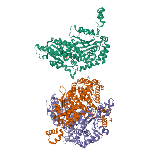
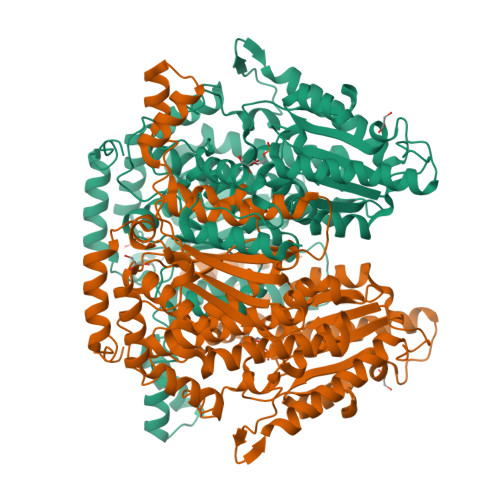
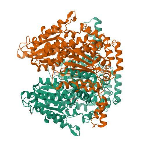
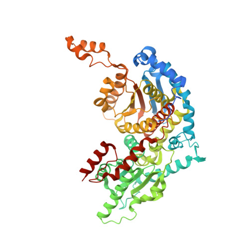
Crystal structure of phosphoglucose isomerase from T. brucei containing glucose-6-phosphate in the active site


Experimental Data Snapshot

  • Method: X-RAY DIFFRACTION
  • Resolution: 1.58 Å
  • R-Value Free: 0.213 
  • R-Value Work: 0.200 

Starting Model: experimental
View more details

wwPDB Validation   3D Report Full Report


Ligand Structure Quality Assessment 


This is version 1.4 of the entry. See complete history


Literature

Crystal structure of phosphoglucose isomerase from Trypanosoma brucei complexed with glucose-6-phosphate at 1.6 A resolution

Arsenieva, D.Appavu, B.L.Mazock, G.H.Jeffery, C.J.

(2008) Proteins 74: 72-80

  • DOI: https://doi.org/10.1002/prot.22133
  • Primary Citation of Related Structures:  
    2O2C, 2O2D

  • PubMed Abstract: 

    Enzymes of glycolysis in Trypanosoma brucei have been identified as potential drug targets for African sleeping sickness because glycolysis is the only source of ATP for the bloodstream form of this parasite. Several inhibitors were previously reported to bind preferentially to trypanosomal phosphoglucose isomerase (PGI, the second enzyme in glycolysis) than to mammalian PGIs, which suggests that PGI might make a good target for species-specific drug design. Herein, we report recombinant expression, purification, crystallization and X-ray crystal structure determination of T. brucei PGI. One structure solved at 1.6 A resolution contains a substrate, D-glucose-6-phosphate, in an extended conformation in the active site. A second structure solved at 1.9 A resolution contains a citrate molecule in the active site. The structures are compared with the crystal structures of PGI from humans and from Leishmania mexicana. The availability of recombinant tPGI and its first high-resolution crystal structures are initial steps in considering this enzyme as a potential drug target.


  • Organizational Affiliation

    Laboratory for Molecular Biology, Department of Biological Sciences, University of Illinois, Chicago, Illinois 60607, USA.


Macromolecules
Find similar proteins by:  (by identity cutoff)  |  3D Structure
Entity ID: 1
MoleculeChains Sequence LengthOrganismDetailsImage
Glucose-6-phosphate isomerase, glycosomal
A, B, C
613Trypanosoma brucei bruceiMutation(s): 0 
Gene Names: PGI
EC: 5.3.1.9
UniProt
Find proteins for P13377 (Trypanosoma brucei brucei)
Explore P13377 
Go to UniProtKB:  P13377
Entity Groups  
Sequence Clusters30% Identity50% Identity70% Identity90% Identity95% Identity100% Identity
UniProt GroupP13377
Sequence Annotations
Expand
  • Reference Sequence
Small Molecules
Ligands 2 Unique
IDChains Name / Formula / InChI Key2D Diagram3D Interactions
G6Q
Query on G6Q

Download Ideal Coordinates CCD File 
D [auth A],
G [auth B],
K [auth C]
GLUCOSE-6-PHOSPHATE
C6 H13 O9 P
VFRROHXSMXFLSN-SLPGGIOYSA-N
GOL
Query on GOL

Download Ideal Coordinates CCD File 
E [auth A]
F [auth A]
H [auth B]
I [auth B]
J [auth B]
E [auth A],
F [auth A],
H [auth B],
I [auth B],
J [auth B],
L [auth C]
GLYCEROL
C3 H8 O3
PEDCQBHIVMGVHV-UHFFFAOYSA-N
Experimental Data & Validation

Experimental Data

  • Method: X-RAY DIFFRACTION
  • Resolution: 1.58 Å
  • R-Value Free: 0.213 
  • R-Value Work: 0.200 
  • Space Group: C 2 2 21
Unit Cell:
Length ( Å )Angle ( ˚ )
a = 125.837α = 90
b = 221.253β = 90
c = 128.087γ = 90
Software Package:
Software NamePurpose
CNSrefinement
SCALEPACKdata scaling
MOLREPphasing
REFMACrefinement
PDB_EXTRACTdata extraction
HKL-2000data collection
HKL-2000data reduction
HKL-2000data scaling
DENZOdata reduction

Structure Validation

View Full Validation Report


Ligand Structure Quality Assessment 


Entry History 

Deposition Data

Revision History  (Full details and data files)

  • Version 1.0: 2007-11-13
    Type: Initial release
  • Version 1.1: 2011-07-13
    Changes: Non-polymer description, Version format compliance
  • Version 1.2: 2017-10-18
    Changes: Refinement description
  • Version 1.3: 2019-07-24
    Changes: Data collection, Refinement description
  • Version 1.4: 2023-08-30
    Changes: Data collection, Database references, Derived calculations, Refinement description