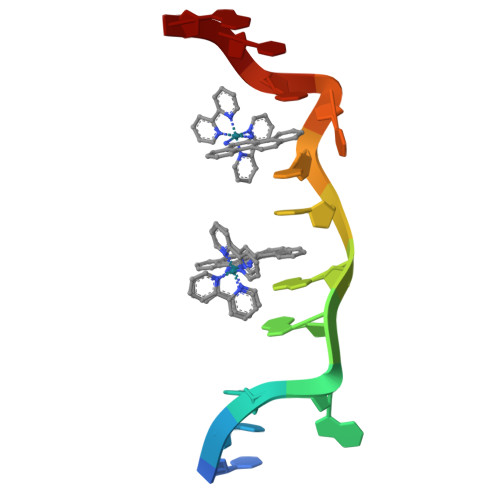
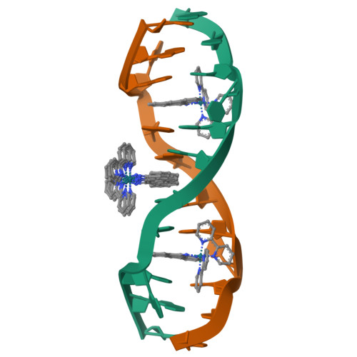
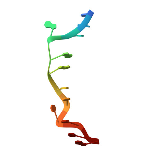
Insights into finding a mismatch through the structure of a mispaired DNA bound by a rhodium intercalator.
Pierre, V.C., Kaiser, J.T., Barton, J.K.(2007) Proc Natl Acad Sci U S A 104: 429-434
- PubMed: 17194756
- DOI: https://doi.org/10.1073/pnas.0610170104
- Primary Citation of Related Structures:
2O1I - PubMed Abstract:
We report the 1.1-A resolution crystal structure of a bulky rhodium complex bound to two different DNA sites, mismatched and matched in the oligonucleotide 5'-(dCGGAAATTCCCG)2-3'. At the AC mismatch site, the structure reveals ligand insertion from the minor groove with ejection of both mismatched bases and elucidates how destabilized mispairs in DNA may be recognized. This unique binding mode contrasts with major groove intercalation, observed at a matched site, where doubling of the base pair rise accommodates stacking of the intercalator. Mass spectral analysis reveals different photocleavage products associated with the two binding modes in the crystal, with only products characteristic of mismatch binding in solution. This structure, illustrating two clearly distinct binding modes for a molecule with DNA, provides a rationale for the interrogation and detection of mismatches.
Organizational Affiliation:
Division of Chemistry and Chemical Engineering, California Institute of Technology, Pasadena, CA 91125, USA.


