2O0A | pdb_00002o0a

The structure of the C-terminal domain of Vik1 has a motor domain fold but lacks a nucleotide-binding site.


Experimental Data Snapshot

  • Method: X-RAY DIFFRACTION
  • Resolution: 1.60 Å
  • R-Value Free: 
    0.261 (Depositor), 0.260 (DCC) 
  • R-Value Work: 
    0.210 (Depositor), 0.208 (DCC) 
  • R-Value Observed: 
    0.212 (Depositor) 

wwPDB Validation 3D Report Full Report

Validation slider image for 2O0A

This is version 1.4 of the entry. See complete history

Literature

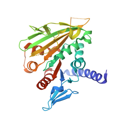
Vik1 modulates microtubule-Kar3 interactions through a motor domain that lacks an active site.

Allingham, J.S.Sproul, L.R.Rayment, I.Gilbert, S.P.

(2007) Cell 128: 1161-1172

  • DOI: https://doi.org/10.1016/j.cell.2006.12.046
  • Primary Citation Related Structures: 
    2O0A

  • PubMed Abstract: 

    Conventional kinesin and class V and VI myosins coordinate the mechanochemical cycles of their motor domains for processive movement of cargo along microtubules or actin filaments. It is widely accepted that this coordination is achieved by allosteric communication or mechanical strain between the motor domains, which controls the nucleotide state and interaction with microtubules or actin. However, questions remain about the interplay between the strain and the nucleotide state. We present an analysis of Saccharomyces cerevisiae Kar3/Vik1, a heterodimeric C-terminal Kinesin-14 containing catalytic Kar3 and the nonmotor protein Vik1. The X-ray crystal structure of Vik1 exhibits a similar fold to the kinesin and myosin catalytic head, but lacks an ATP binding site. Vik1 binds more tightly to microtubules than Kar3 and facilitates cooperative microtubule decoration by Kar3/Vik1 heterodimers, and yet allows motility. These results demand communication between Vik1 and Kar3 via a mechanism that coordinates their interactions with microtubules.


  • Organizational Affiliation
    • Department of Biochemistry, University of Wisconsin, Madison, WI 53706, USA.

Macromolecule Content 

  • Total Structure Weight: 34.69 kDa 
  • Atom Count: 2,454 
  • Modeled Residue Count: 270 
  • Deposited Residue Count: 298 
  • Unique protein chains: 1

Macromolecules

Find similar proteins by:|  3D Structure
Entity ID: 1
MoleculeChains  Sequence LengthOrganismDetailsImage
S.cerevisiae chromosome XVI reading frame ORF YPL253c298Saccharomyces cerevisiaeMutation(s): 0 
UniProt
Find proteins for Q12045 (Saccharomyces cerevisiae (strain ATCC 204508 / S288c))
Explore Q12045 
Go to UniProtKB:  Q12045
Entity Groups
Sequence Clusters30% Identity50% Identity70% Identity90% Identity95% Identity100% Identity
UniProt GroupQ12045
Sequence Annotations
Expand
Reference Sequence

Small Molecules

Ligands 1 Unique
IDChains Name / Formula / InChI Key2D Diagram3D Interactions
EDO

Query on EDO


Download:Ideal Coordinates CCD File
B [auth A]1,2-ETHANEDIOL
C2 H6 O2
LYCAIKOWRPUZTN-UHFFFAOYSA-N

Experimental Data & Validation

Experimental Data

  • Method: X-RAY DIFFRACTION
  • Resolution: 1.60 Å
  • R-Value Free:  0.261 (Depositor), 0.260 (DCC) 
  • R-Value Work:  0.210 (Depositor), 0.208 (DCC) 
  • R-Value Observed: 0.212 (Depositor) 
Space Group: P 21 21 21
Unit Cell:
Length ( Å )Angle ( ˚ )
a = 46.524α = 90
b = 70.129β = 90
c = 79.861γ = 90
Software Package:
Software NamePurpose
DENZOdata reduction
SCALEPACKdata scaling
SOLVEphasing
RESOLVEphasing
REFMACrefinement
PDB_EXTRACTdata extraction
HKL-2000data collection
HKL-2000data reduction

Structure Validation

View Full Validation Report


Entry History 

Deposition Data

Revision History  (Full details and data files)

  • Version 1.0: 2007-03-27
    Type: Initial release
  • Version 1.1: 2008-05-01
    Changes: Version format compliance
  • Version 1.2: 2011-07-13
    Changes: Version format compliance
  • Version 1.3: 2017-10-18
    Changes: Refinement description
  • Version 1.4: 2023-12-27
    Changes: Data collection, Database references, Derived calculations