2NZX | pdb_00002nzx

Crystal Structure of alpha1,3-Fucosyltransferase with GDP


Experimental Data Snapshot

  • Method: X-RAY DIFFRACTION
  • Resolution: 1.90 Å
  • R-Value Free: 
    0.237 (Depositor), 0.237 (DCC) 
  • R-Value Work: 
    0.190 (Depositor), 0.190 (DCC) 
  • R-Value Observed: 
    0.192 (Depositor) 

wwPDB Validation 3D Report Full Report

Validation slider image for 2NZX

Ligand Structure Quality Assessment 


This is version 1.4 of the entry. See complete history

Literature

Structure and mechanism of Helicobacter pylori fucosyltransferase. A basis for lipopolysaccharide variation and inhibitor design.

Sun, H.Y.Lin, S.W.Ko, T.P.Pan, J.F.Liu, C.L.Lin, C.N.Wang, A.H.Lin, C.H.

(2007) J Biological Chem 282: 9973-9982

  • DOI: https://doi.org/10.1074/jbc.M610285200
  • Primary Citation Related Structures: 
    2NZW, 2NZX, 2NZY

  • PubMed Abstract: 

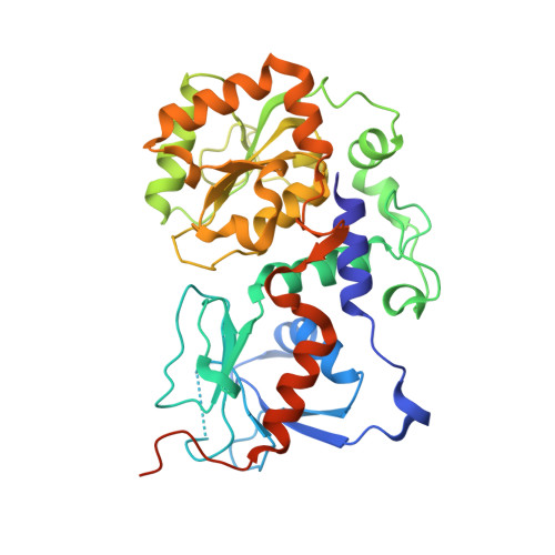
    Helicobacter pylori alpha1,3-fucosyltransferase (FucT) is involved in catalysis to produce the Lewis x trisaccharide, the major component of the bacteria's lipopolysaccharides, which has been suggested to mimic the surface sugars in gastric epithelium to escape host immune surveillance. We report here three x-ray crystal structures of FucT, including the FucT.GDP-fucose and FucT.GDP complexes. The protein structure is typical of the glycosyltransferase-B family despite little sequence homology. We identified a number of catalytically important residues, including Glu-95, which serves as the general base, and Glu-249, which stabilizes the developing oxonium ion during catalysis. The residues Arg-195, Tyr-246, Glu-249, and Lys-250 serve to interact with the donor substrate, GDP-fucose. Variations in the protein and ligand conformations, as well as a possible FucT dimer, were also observed. We propose a catalytic mechanism and a model of polysaccharide binding not only to explain the observed variations in H. pylori lipopolysaccharides, but also to facilitate the development of potent inhibitors.


  • Organizational Affiliation
    • Institute of Biochemical Sciences, National Taiwan University, Taipei 10642; Institute of Biological Chemistry, Academia Sinica, No.128 Academia Road Section 2, Nan-Kang, Taipei 11529, Taiwan.

Macromolecule Content 

  • Total Structure Weight: 130.8 kDa 
  • Atom Count: 10,752 
  • Modeled Residue Count: 1,028 
  • Deposited Residue Count: 1,113 
  • Unique protein chains: 1

Macromolecules

Find similar proteins by:|  3D Structure
Entity ID: 1
MoleculeChains  Sequence LengthOrganismDetailsImage
Alpha1,3-fucosyltransferase
A, B, C
371Helicobacter pyloriMutation(s): 0 
Gene Names: AF008596
EC: 2.4.1.152
UniProt
Find proteins for O30511 (Helicobacter pylori)
Explore O30511 
Go to UniProtKB:  O30511
Entity Groups
Sequence Clusters30% Identity50% Identity70% Identity90% Identity95% Identity100% Identity
UniProt GroupO30511
Sequence Annotations
Expand
Reference Sequence

Experimental Data & Validation

Experimental Data

  • Method: X-RAY DIFFRACTION
  • Resolution: 1.90 Å
  • R-Value Free:  0.237 (Depositor), 0.237 (DCC) 
  • R-Value Work:  0.190 (Depositor), 0.190 (DCC) 
  • R-Value Observed: 0.192 (Depositor) 
Space Group: P 21 21 2
Unit Cell:
Length ( Å )Angle ( ˚ )
a = 104.154α = 90
b = 136.249β = 90
c = 96.375γ = 90
Software Package:
Software NamePurpose
HKL-2000data collection
CNSrefinement
HKL-2000data reduction
HKL-2000data scaling
CNSphasing

Structure Validation

View Full Validation Report


Ligand Structure Quality Assessment 


Entry History 

Deposition Data

Revision History  (Full details and data files)

  • Version 1.0: 2007-01-23
    Type: Initial release
  • Version 1.1: 2008-05-01
    Changes: Version format compliance
  • Version 1.2: 2011-07-13
    Changes: Version format compliance
  • Version 1.3: 2017-07-05
    Changes: Database references
  • Version 1.4: 2023-12-27
    Changes: Data collection, Database references, Derived calculations