2NW0 | pdb_00002nw0

Crystal structure of a lysin


Experimental Data Snapshot

  • Method: X-RAY DIFFRACTION
  • Resolution: 1.60 Å
  • R-Value Free: 
    0.195 (Depositor), 0.204 (DCC) 
  • R-Value Work: 
    0.149 (Depositor), 0.161 (DCC) 
  • R-Value Observed: 
    0.151 (Depositor) 

Starting Model: experimental
View more details

wwPDB Validation 3D Report Full Report

Validation slider image for 2NW0

This is version 1.3 of the entry. See complete history

Literature

The 1.6 A Crystal Structure of the Catalytic Domain of PlyB, a Bacteriophage Lysin Active Against Bacillus anthracis.

Porter, C.J.Schuch, R.Pelzek, A.J.Buckle, A.M.McGowan, S.Wilce, M.C.Rossjohn, J.Russell, R.Nelson, D.Fischetti, V.A.Whisstock, J.C.

(2007) J Mol Biology 366: 540-550

  • DOI: https://doi.org/10.1016/j.jmb.2006.11.056
  • Primary Citation Related Structures: 
    2NW0

  • PubMed Abstract: 

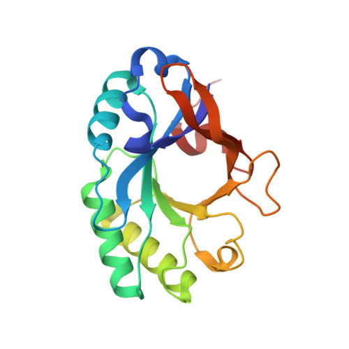
    Lysins are peptidoglycan hydrolases that are produced by bacteriophage and act to lyse the bacterial host cell wall during progeny phage release. Here, we describe the structure and function of a novel bacteriophage-derived lysin, PlyB, which displays potent lytic activity against the Bacillus anthracis-like strain ATCC 4342. This molecule comprises an N-terminal catalytic domain (PlyB(cat)) and a C-terminal bacterial SH3-like domain, SH3b. It is shown that both domains are required for effective catalytic activity against ATCC 4342. Further, PlyB has specific activity comparable to the phage lysin PlyG, an amidase being developed as a therapeutic against anthrax. In contrast to PlyG, however, the 1.6 A X-ray crystal structure of PlyB(cat) reveals that the catalytic domain adopts the glycosyl hydrolase (GH)-25, rather than phage T7 lysozyme-like fold. PlyB therefore represents a new class of anthrax lysin and a new defensive tool in the armament against anthrax-mediated bioterrorism.


  • Organizational Affiliation
    • Protein Crystallography Unit, Department of Biochemistry and Molecular Biology and The ARC Centre of Excellence for Structural and Functional Microbial Genomics, Monash University, Clayton, Melbourne, VIC 3800, Australia.

Macromolecule Content 

  • Total Structure Weight: 43.39 kDa 
  • Atom Count: 3,581 
  • Modeled Residue Count: 378 
  • Deposited Residue Count: 378 
  • Unique protein chains: 1

Macromolecules

Find similar proteins by:|  3D Structure
Entity ID: 1
MoleculeChains  Sequence LengthOrganismDetailsImage
PlyB
A, B
189Bacteriophage sp.Mutation(s): 0 
Gene Names: PlyB
EC: 3.2.1.17

Experimental Data & Validation

Experimental Data

  • Method: X-RAY DIFFRACTION
  • Resolution: 1.60 Å
  • R-Value Free:  0.195 (Depositor), 0.204 (DCC) 
  • R-Value Work:  0.149 (Depositor), 0.161 (DCC) 
  • R-Value Observed: 0.151 (Depositor) 
Space Group: P 1 21 1
Unit Cell:
Length ( Å )Angle ( ˚ )
a = 37.055α = 90
b = 65.773β = 85.08
c = 66.747γ = 90
Software Package:
Software NamePurpose
SCALAdata scaling
MOLREPphasing
REFMACrefinement
PDB_EXTRACTdata extraction
CrystalCleardata collection
MOSFLMdata reduction
CCP4data scaling

Structure Validation

View Full Validation Report


Entry History 

Deposition Data

Revision History  (Full details and data files)

  • Version 1.0: 2006-12-05
    Type: Initial release
  • Version 1.1: 2008-05-01
    Changes: Version format compliance
  • Version 1.2: 2011-07-13
    Changes: Advisory, Version format compliance
  • Version 1.3: 2023-10-25
    Changes: Data collection, Database references, Derived calculations, Refinement description