2NUE

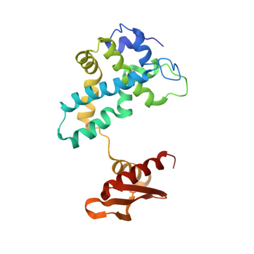
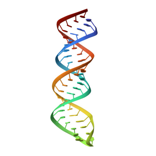
Crystal structure of RNase III from Aquifex aeolicus complexed with ds-RNA at 2.9-Angstrom Resolution


Experimental Data Snapshot

  • Method: X-RAY DIFFRACTION
  • Resolution: 2.90 Å
  • R-Value Free: 0.275 
  • R-Value Work: 0.238 
  • R-Value Observed: 0.238 

Starting Model: experimental
View more details

wwPDB Validation   3D Report Full Report


This is version 1.3 of the entry. See complete history


Literature

A stepwise model for double-stranded RNA processing by ribonuclease III.

Gan, J.Shaw, G.Tropea, J.E.Waugh, D.S.Court, D.L.Ji, X.

(2007) Mol Microbiol 67: 143-154

  • DOI: https://doi.org/10.1111/j.1365-2958.2007.06032.x
  • Primary Citation of Related Structures:  
    2NUE, 2NUF, 2NUG

  • PubMed Abstract: 

    RNA interference is mediated by small interfering RNAs produced by members of the ribonuclease III (RNase III) family represented by bacterial RNase III and eukaryotic Rnt1p, Drosha and Dicer. For mechanistic studies, bacterial RNase III has been a valuable model system for the family. Previously, we have shown that RNase III uses two catalytic sites to create the 2-nucleotide (nt) 3' overhangs in its products. Here, we present three crystal structures of RNase III in complex with double-stranded RNA, demonstrating how Mg(2+) is essential for the formation of a catalytically competent protein-RNA complex, how the use of two Mg(2+) ions can drive the hydrolysis of each phosphodiester bond, and how conformational changes in both the substrate and the protein are critical elements for assembling the catalytic complex. Moreover, we have modelled a protein-substrate complex and a protein-reaction intermediate (transition state) complex on the basis of the crystal structures. Together, the crystal structures and the models suggest a stepwise mechanism for RNase III to execute the phosphoryl transfer reaction.


  • Organizational Affiliation

    Center for Cancer Research, National Cancer Institute, National Institutes of Health, Frederick, MD 21702, USA.


Macromolecules

Find similar proteins by:  (by identity cutoff)  |  3D Structure
Entity ID: 2
MoleculeChains Sequence LengthOrganismDetailsImage
Ribonuclease IIIB [auth A],
C [auth B]
221Aquifex aeolicusMutation(s): 0 
Gene Names: rnc
EC: 3.1.26.3
UniProt
Find proteins for O67082 (Aquifex aeolicus (strain VF5))
Explore O67082 
Go to UniProtKB:  O67082
Entity Groups  
Sequence Clusters30% Identity50% Identity70% Identity90% Identity95% Identity100% Identity
UniProt GroupO67082
Sequence Annotations
Expand
  • Reference Sequence
Find similar nucleic acids by:  (by identity cutoff)  |  3D Structure
Entity ID: 1
MoleculeChains LengthOrganismImage
46-MERA [auth C]46N/A
Sequence Annotations
Expand
  • Reference Sequence
Experimental Data & Validation

Experimental Data

  • Method: X-RAY DIFFRACTION
  • Resolution: 2.90 Å
  • R-Value Free: 0.275 
  • R-Value Work: 0.238 
  • R-Value Observed: 0.238 
  • Space Group: C 1 2 1
Unit Cell:
Length ( Å )Angle ( ˚ )
a = 101.596α = 90
b = 101.657β = 103.69
c = 67.082γ = 90
Software Package:
Software NamePurpose
CNSrefinement
MAR345data collection
HKL-2000data reduction
HKL-2000data scaling
PHASERphasing

Structure Validation

View Full Validation Report


Entry History 

Deposition Data

Revision History  (Full details and data files)

  • Version 1.0: 2007-11-20
    Type: Initial release
  • Version 1.1: 2011-07-13
    Changes: Version format compliance
  • Version 1.2: 2017-10-18
    Changes: Refinement description
  • Version 1.3: 2023-08-30
    Changes: Data collection, Database references, Refinement description, Structure summary