Structural basis for activation of the therapeutic L-nucleoside analogs 3TC and troxacitabine by human deoxycytidine kinase.
Sabini, E., Hazra, S., Konrad, M., Burley, S.K., Lavie, A.(2007) Nucleic Acids Res 35: 186-192
- PubMed: 17158155 Search on PubMedSearch on PubMed Central
- DOI: https://doi.org/10.1093/nar/gkl1038
- Primary Citation Related Structures:
2NO9, 2NOA - PubMed Abstract:
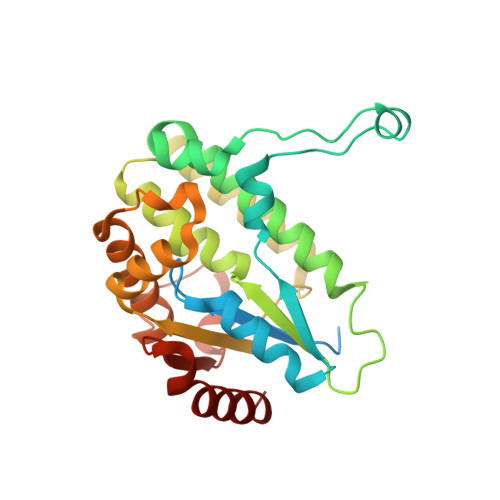
L-nucleoside analogs represent an important class of small molecules for treating both viral infections and cancers. These pro-drugs achieve pharmacological activity only after enzyme-catalyzed conversion to their tri-phosphorylated forms. Herein, we report the crystal structures of human deoxycytidine kinase (dCK) in complex with the L-nucleosides (-)-beta-2',3'-dideoxy-3'-thiacytidine (3TC)--an approved anti-human immunodeficiency virus (HIV) agent--and troxacitabine (TRO)--an experimental anti-neoplastic agent. The first step in activating these agents is catalyzed by dCK. Our studies reveal how dCK, which normally catalyzes phosphorylation of the natural D-nucleosides, can efficiently phosphorylate substrates with non-physiologic chirality. The capability of dCK to phosphorylate both D- and L-nucleosides and nucleoside analogs derives from structural properties of both the enzyme and the substrates themselves. First, the nucleoside-binding site tolerates substrates with different chiral configurations by maintaining virtually all of the protein-ligand interactions responsible for productive substrate positioning. Second, the pseudo-symmetry of nucleosides and nucleoside analogs in combination with their conformational flexibility allows the L- and D-enantiomeric forms to adopt similar shapes when bound to the enzyme. This is the first analysis of the structural basis for activation of L-nucleoside analogs, providing further impetus for discovery and clinical development of new agents in this molecular class.
- Department of Biochemistry and Molecular Genetics, University of Illinois Chicago, 900 S. Ashland (M/C 669), Chicago, IL 60607, USA.
Organizational Affiliation: