2NMT | pdb_00002nmt

MYRISTOYL-COA:PROTEIN N-MYRISTOYLTRANSFERASE BOUND TO MYRISTOYL-COA AND PEPTIDE ANALOGS


Experimental Data Snapshot

  • Method: X-RAY DIFFRACTION
  • Resolution: 2.90 Å
  • R-Value Free: 
    0.297 (Depositor) 
  • R-Value Work: 
    0.228 (Depositor) 
  • R-Value Observed: 
    0.228 (Depositor) 

wwPDB Validation 3D Report Full Report

Validation slider image for 2NMT

This is version 1.3 of the entry. See complete history

Literature

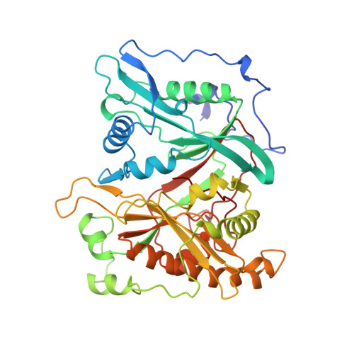
Structure of N-myristoyltransferase with bound myristoylCoA and peptide substrate analogs.

Bhatnagar, R.S.Futterer, K.Farazi, T.A.Korolev, S.Murray, C.L.Jackson-Machelski, E.Gokel, G.W.Gordon, J.I.Waksman, G.

(1998) Nat Struct Biol 5: 1091-1097

  • DOI: https://doi.org/10.1038/4202
  • Primary Citation Related Structures: 
    2NMT

  • PubMed Abstract: 

    N-myristoyltransferase (Nmt) attaches myristate to the N-terminal glycine of many important eukaryotic and viral proteins. It is a target for anti-fungal and anti-viral therapy. We have determined the structure, to 2.9 A resolution, of a ternary complex of Saccharomyces cerevisiae Nmt1p with bound myristoylCoA and peptide substrate analogs. The model reveals structural features that define the enzyme's substrate specificities and regulate the ordered binding and release of substrates and products. A novel catalytic mechanism is proposed involving deprotonation of the N-terminal ammonium of a peptide substrate by the enzyme's C-terminal backbone carboxylate.


  • Organizational Affiliation
    • Department of Molecular Biology and Pharmacology, Washington University School of Medicine, St. Louis, Missouri 63110, USA.

Macromolecule Content 

  • Total Structure Weight: 51.71 kDa 
  • Atom Count: 3,601 
  • Modeled Residue Count: 422 
  • Deposited Residue Count: 422 
  • Unique protein chains: 1

Macromolecules

Find similar proteins by:|  3D Structure
Entity ID: 1
MoleculeChains  Sequence LengthOrganismDetailsImage
MYRISTOYL-COA\:PROTEIN N-MYRISTOYLTRANSFERASE422Saccharomyces cerevisiaeMutation(s): 0 
Gene Names: NMT1
EC: 2.3.1.97
UniProt
Find proteins for P14743 (Saccharomyces cerevisiae (strain ATCC 204508 / S288c))
Explore P14743 
Go to UniProtKB:  P14743
Entity Groups
Sequence Clusters30% Identity50% Identity70% Identity90% Identity95% Identity100% Identity
UniProt GroupP14743
Sequence Annotations
Expand
Reference Sequence

Small Molecules

Ligands 3 Unique
IDChains Name / Formula / InChI Key2D Diagram3D Interactions
NHM

Query on NHM


Download:Ideal Coordinates CCD File
B [auth A]S-(2-OXO)PENTADECYLCOA
C36 H64 N7 O17 P3 S
JKWHUJMJVNMKEF-SXKLBCNJSA-N
MIM

Query on MIM


Download:Ideal Coordinates CCD File
C [auth A][CYCLOHEXYLETHYL]-[[[[4-[2-METHYL-1-IMIDAZOLYL-BUTYL]PHENYL]ACETYL]-SERYL]-LYSINYL]-AMINE
C33 H52 N6 O4
WHLPIOSHBKQGHA-KYJUHHDHSA-N
GOL

Query on GOL


Download:Ideal Coordinates CCD File
D [auth A]
E [auth A]
F [auth A]
G [auth A]
H [auth A]
D [auth A],
E [auth A],
F [auth A],
G [auth A],
H [auth A],
I [auth A],
J [auth A],
K [auth A],
L [auth A],
M [auth A],
N [auth A],
O [auth A]
GLYCEROL
C3 H8 O3
PEDCQBHIVMGVHV-UHFFFAOYSA-N

Experimental Data & Validation

Experimental Data

  • Method: X-RAY DIFFRACTION
  • Resolution: 2.90 Å
  • R-Value Free:  0.297 (Depositor) 
  • R-Value Work:  0.228 (Depositor) 
  • R-Value Observed: 0.228 (Depositor) 
Space Group: P 31 2 1
Unit Cell:
Length ( Å )Angle ( ˚ )
a = 105.4α = 90
b = 105.4β = 90
c = 106.7γ = 120
Software Package:
Software NamePurpose
CNSrefinement
SCALEPACKdata scaling
CNSphasing

Structure Validation

View Full Validation Report


Entry History 

Deposition Data

Revision History  (Full details and data files)

  • Version 1.0: 1999-01-06
    Type: Initial release
  • Version 1.1: 2008-03-24
    Changes: Version format compliance
  • Version 1.2: 2011-07-13
    Changes: Non-polymer description, Version format compliance
  • Version 1.3: 2024-12-25
    Changes: Advisory, Data collection, Database references, Derived calculations, Structure summary