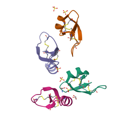
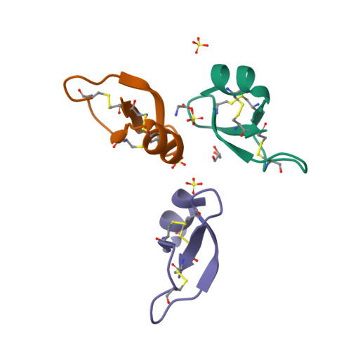
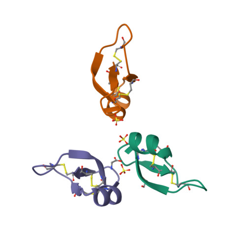
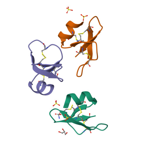
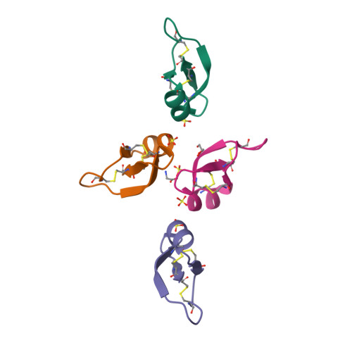
2NLQ

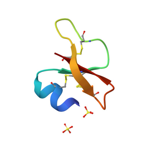
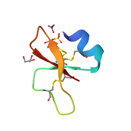
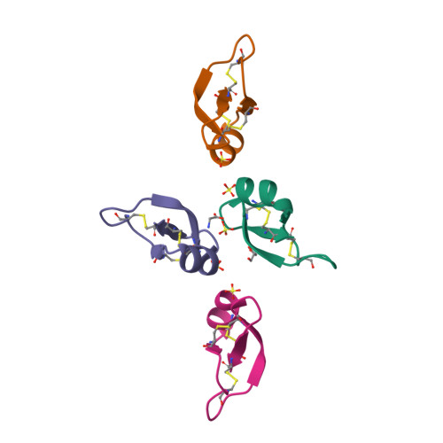
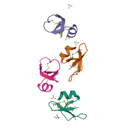
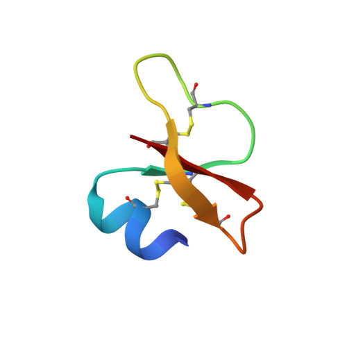
Human beta-defensin-1 (Mutant Lys31Ala)


Experimental Data Snapshot

  • Method: X-RAY DIFFRACTION
  • Resolution: 1.80 Å
  • R-Value Free: 0.257 
  • R-Value Work: 0.179 
  • R-Value Observed: 0.183 

Starting Model: experimental
View more details

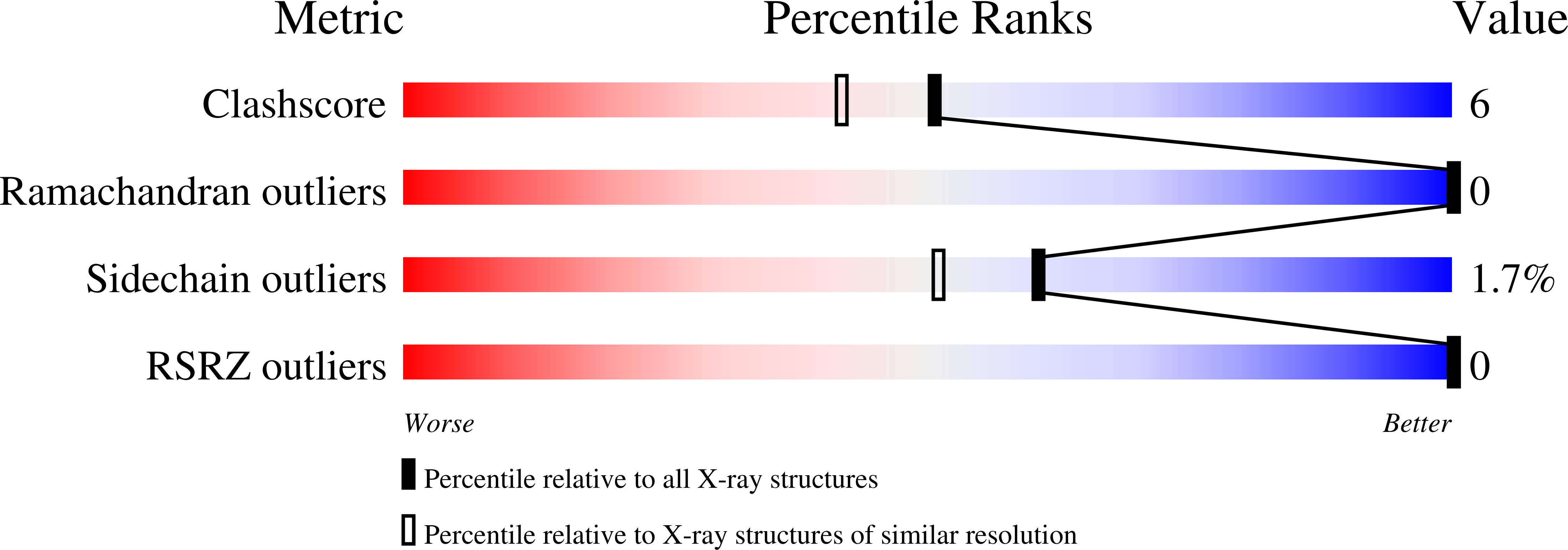
wwPDB Validation   3D Report Full Report


This is version 1.6 of the entry. See complete history


Literature

Studies of the Biological Properties of Human beta-Defensin 1.

Pazgier, M.Prahl, A.Hoover, D.M.Lubkowski, J.

(2007) J Biol Chem 282: 1819-1829

  • DOI: https://doi.org/10.1074/jbc.M607210200
  • Primary Citation of Related Structures:  
    2NLB, 2NLC, 2NLD, 2NLE, 2NLF, 2NLG, 2NLH, 2NLP, 2NLQ, 2NLS

  • PubMed Abstract: 

    Defensins are small (30-45 amino acid residues) cationic proteins with broad antimicrobial activity against many bacteria and fungi, some enveloped viruses, and other activities such as chemoattraction of a range of different cell types to the sites of inflammation. These proteins represent attractive targets for developing novel antimicrobial agents and modulators of immune responses with therapeutic applicability. In this report, we present the results of functional and structural studies of 26 single-site mutants of human beta-defensin 1 (hBD1). All mutants were assayed for antimicrobial activity against Escherichia coli (ATCC strain 25922) and for chemotactic activity with CCR6-transfected HEK293 cells. To analyze the structural implications of mutagenesis and to verify the correctness of the disulfide connectivity, we used x-ray crystallography to conduct complete structural studies for 10 mutants in which the topology of disulfides was the same as in the native hBD1. Mutations did not induce significant changes of the tertiary structure, suggesting that the observed alterations of biological properties of the mutants were solely associated with changes in the respective side chains. We found that cationic residues located near the C terminus (Arg(29), Lys(31), Lys(33), and Lys(36)) of hBD1 define most of the anti-E. coli in vitro activity of this protein. In turn, nearly all mutations altering the CCR6-mediated chemotaxis are located at one area of the protein, defined by the N-terminal alpha-helical region (Asp(1)... Ser(8)) and a few topologically adjacent residues (Lys(22), Arg(29), and Lys(33)). These experimental results allow for the first time drafting of the CCR6-epitope for a defensin molecule.


  • Organizational Affiliation

    Macromolecular Crystallography Laboratory, NCI, National Institutes of Health, Frederick, Maryland 21702, USA.


Macromolecules
Find similar proteins by:  (by identity cutoff)  |  3D Structure
Entity ID: 1
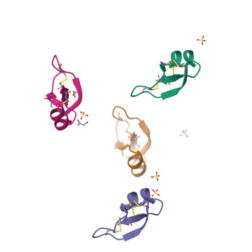
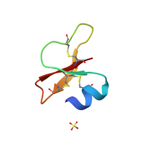
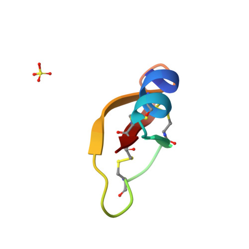
MoleculeChains Sequence LengthOrganismDetailsImage
Beta-defensin 1
A, B, C, D
36Homo sapiensMutation(s): 1 
Gene Names: DEFB1BD1HBD1
UniProt & NIH Common Fund Data Resources
Find proteins for P60022 (Homo sapiens)
Explore P60022 
Go to UniProtKB:  P60022
PHAROS:  P60022
GTEx:  ENSG00000164825 
Entity Groups  
Sequence Clusters30% Identity50% Identity70% Identity90% Identity95% Identity100% Identity
UniProt GroupP60022
Sequence Annotations
Expand
  • Reference Sequence
Small Molecules
Ligands 3 Unique
IDChains Name / Formula / InChI Key2D Diagram3D Interactions
SO4
Query on SO4

Download Ideal Coordinates CCD File 
E [auth A],
F [auth B],
G [auth C],
H [auth C],
I [auth D]
SULFATE ION
O4 S
QAOWNCQODCNURD-UHFFFAOYSA-L
GOL
Query on GOL

Download Ideal Coordinates CCD File 
K [auth D]GLYCEROL
C3 H8 O3
PEDCQBHIVMGVHV-UHFFFAOYSA-N
GLY
Query on GLY

Download Ideal Coordinates CCD File 
J [auth D]GLYCINE
C2 H5 N O2
DHMQDGOQFOQNFH-UHFFFAOYSA-N
Experimental Data & Validation

Experimental Data

  • Method: X-RAY DIFFRACTION
  • Resolution: 1.80 Å
  • R-Value Free: 0.257 
  • R-Value Work: 0.179 
  • R-Value Observed: 0.183 
  • Space Group: C 1 2 1
Unit Cell:
Length ( Å )Angle ( ˚ )
a = 97.89α = 90
b = 27.68β = 113.5
c = 58.26γ = 90
Software Package:
Software NamePurpose
DENZOdata reduction
SCALEPACKdata scaling
REFMACrefinement
PDB_EXTRACTdata extraction
MAR345data collection
HKL-2000data reduction
HKL-2000data scaling
AMoREphasing

Structure Validation

View Full Validation Report


Entry History 

Deposition Data

Revision History  (Full details and data files)

  • Version 1.0: 2006-10-31
    Type: Initial release
  • Version 1.1: 2008-05-01
    Changes: Version format compliance
  • Version 1.2: 2011-07-13
    Changes: Non-polymer description, Version format compliance
  • Version 1.3: 2017-10-18
    Changes: Refinement description
  • Version 1.4: 2021-10-20
    Changes: Database references, Derived calculations
  • Version 1.5: 2023-08-30
    Changes: Data collection, Refinement description
  • Version 1.6: 2024-10-30
    Changes: Structure summary