2NLA | pdb_00002nla

Crystal structure of the Mcl-1:mNoxaB BH3 complex


Experimental Data Snapshot

  • Method: X-RAY DIFFRACTION
  • Resolution: 2.80 Å
  • R-Value Free: 
    0.291 (Depositor), 0.308 (DCC) 
  • R-Value Work: 
    0.207 (Depositor), 0.261 (DCC) 
  • R-Value Observed: 
    0.211 (Depositor) 

wwPDB Validation 3D Report Full Report

Validation slider image for 2NLA

This is version 1.6 of the entry. See complete history

Literature

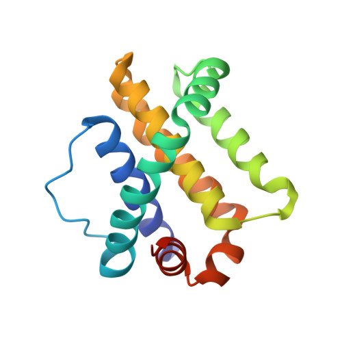
Structural insights into the degradation of Mcl-1 induced by BH3 domains.

Czabotar, P.E.Lee, E.F.van Delft, M.F.Day, C.L.Smith, B.J.Huang, D.C.Fairlie, W.D.Hinds, M.G.Colman, P.M.

(2007) Proc Natl Acad Sci U S A 104: 6217-6222

  • DOI: https://doi.org/10.1073/pnas.0701297104
  • Primary Citation Related Structures: 
    2JM6, 2NL9, 2NLA

  • PubMed Abstract: 

    Apoptosis is held in check by prosurvival proteins of the Bcl-2 family. The distantly related BH3-only proteins bind to and antagonize them, thereby promoting apoptosis. Whereas binding of the BH3-only protein Noxa to prosurvival Mcl-1 induces Mcl-1 degradation by the proteasome, binding of another BH3-only ligand, Bim, elevates Mcl-1 protein levels. We compared the three-dimensional structures of the complexes formed between BH3 peptides of both Bim and Noxa, and we show that a discrete C-terminal sequence of the Noxa BH3 is necessary to instigate Mcl-1 degradation.


  • Organizational Affiliation
    • The Walter and Eliza Hall Institute of Medical Research, Parkville, Victoria 3050, Australia.

Macromolecule Content 

  • Total Structure Weight: 20.83 kDa 
  • Atom Count: 1,406 
  • Modeled Residue Count: 171 
  • Deposited Residue Count: 183 
  • Unique protein chains: 2

Macromolecules

Find similar proteins by:|  3D Structure
Entity ID: 1
MoleculeChains  Sequence LengthOrganismDetailsImage
FUSION PROTEIN CONSISTING OF Induced myeloid leukemia cell differentiation protein Mcl-1 homolog157Mus musculusHomo sapiens
This entity is chimeric
Mutation(s): 0 
Gene Names: Mcl1
UniProt & NIH Common Fund Data Resources
Find proteins for P97287 (Mus musculus)
Explore P97287 
Go to UniProtKB:  P97287
IMPC:  MGI:101769
Find proteins for Q07820 (Homo sapiens)
Explore Q07820 
Go to UniProtKB:  Q07820
PHAROS:  Q07820
GTEx:  ENSG00000143384 
Entity Groups
Sequence Clusters30% Identity50% Identity70% Identity90% Identity95% Identity100% Identity
UniProt GroupsP97287Q07820
Sequence Annotations
Expand
Reference Sequence
Find similar proteins by:|  3D Structure
Entity ID: 2
MoleculeChains  Sequence LengthOrganismDetailsImage
Phorbol-12-myristate-13-acetate-induced protein 126Mus musculusMutation(s): 0 
UniProt
Find proteins for Q9JM54 (Mus musculus)
Explore Q9JM54 
Go to UniProtKB:  Q9JM54
Entity Groups
Sequence Clusters30% Identity50% Identity70% Identity90% Identity95% Identity100% Identity
UniProt GroupQ9JM54
Sequence Annotations
Expand
Reference Sequence

Experimental Data & Validation

Experimental Data

  • Method: X-RAY DIFFRACTION
  • Resolution: 2.80 Å
  • R-Value Free:  0.291 (Depositor), 0.308 (DCC) 
  • R-Value Work:  0.207 (Depositor), 0.261 (DCC) 
  • R-Value Observed: 0.211 (Depositor) 
Space Group: P 63
Unit Cell:
Length ( Å )Angle ( ˚ )
a = 85.381α = 90
b = 85.381β = 90
c = 46.916γ = 120
Software Package:
Software NamePurpose
DENZOdata reduction
SCALEPACKdata scaling
REFMACrefinement
PDB_EXTRACTdata extraction
HKL-2000data collection
HKL-2000data reduction
PHASERphasing

Structure Validation

View Full Validation Report


Entry History 

Deposition Data

Revision History  (Full details and data files)

  • Version 1.0: 2007-03-27
    Type: Initial release
  • Version 1.1: 2008-05-01
    Changes: Version format compliance
  • Version 1.2: 2011-07-13
    Changes: Version format compliance
  • Version 1.3: 2017-06-14
    Changes: Source and taxonomy, Structure summary
  • Version 1.4: 2017-10-18
    Changes: Refinement description
  • Version 1.5: 2023-12-27
    Changes: Advisory, Data collection, Database references
  • Version 1.6: 2024-11-20
    Changes: Structure summary