2NG1 | pdb_00002ng1

N AND GTPASE DOMAINS OF THE SIGNAL SEQUENCE RECOGNITION PROTEIN FFH FROM THERMUS AQUATICUS


Experimental Data Snapshot

  • Method: X-RAY DIFFRACTION
  • Resolution: 2.02 Å
  • R-Value Free: 
    0.290 (Depositor) 
  • R-Value Work: 
    0.200 (Depositor), 0.210 (DCC) 
  • R-Value Observed: 
    0.200 (Depositor) 

Starting Model: experimental
View more details

wwPDB Validation 3D Report Full Report

Validation slider image for 2NG1

Ligand Structure Quality Assessment 


This is version 1.5 of the entry. See complete history

Literature

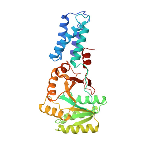
Functional changes in the structure of the SRP GTPase on binding GDP and Mg2+GDP.

Freymann, D.M.Keenan, R.J.Stroud, R.M.Walter, P.

(1999) Nat Struct Biol 6: 793-801

  • DOI: https://doi.org/10.1038/11572
  • Primary Citation Related Structures: 
    1NG1, 2NG1, 3NG1

  • PubMed Abstract: 

    Ffh is a component of a bacterial ribonucleoprotein complex homologous to the signal recognition particle (SRP) of eukaryotes. It comprises three domains that mediate both binding to the hydrophobic signal sequence of the nascent polypeptide and the GTP-dependent interaction of Ffh with a structurally homologous GTPase of the SRP receptor. The X-ray structures of the two-domain 'NG' GTPase of Ffh in complex with Mg2+GDP and GDP have been determined at 2.0 A resolution. The structures explain the low nucleotide affinity of Ffh and locate two regions of structural mobility at opposite sides of the nucleotide-binding site. One of these regions includes highly conserved sequence motifs that presumably contribute to the structural trigger signaling the GTP-bound state. The other includes the highly conserved interface between the N and G domains, and supports the hypothesis that the N domain regulates or signals the nucleotide occupancy of the G domain.


  • Organizational Affiliation
    • Department of Molecular Pharmacology and Biological Chemistry, Northwestern University Medical School, 303 E. Chicago Avenue, Chicago, Illinois 60611, USA. freymann@nwu.edu

Macromolecule Content 

  • Total Structure Weight: 32.85 kDa 
  • Atom Count: 2,375 
  • Modeled Residue Count: 293 
  • Deposited Residue Count: 293 
  • Unique protein chains: 1

Macromolecules

Find similar proteins by:|  3D Structure
Entity ID: 1
MoleculeChains  Sequence LengthOrganismDetailsImage
SIGNAL SEQUENCE RECOGNITION PROTEIN FFH293Thermus aquaticusMutation(s): 1 
Gene Names: FFH
EC: 3.6.5.4
UniProt
Find proteins for O07347 (Thermus aquaticus)
Explore O07347 
Go to UniProtKB:  O07347
Entity Groups
Sequence Clusters30% Identity50% Identity70% Identity90% Identity95% Identity100% Identity
UniProt GroupO07347
Sequence Annotations
Expand
Reference Sequence

Experimental Data & Validation

Experimental Data

  • Method: X-RAY DIFFRACTION
  • Resolution: 2.02 Å
  • R-Value Free:  0.290 (Depositor) 
  • R-Value Work:  0.200 (Depositor), 0.210 (DCC) 
  • R-Value Observed: 0.200 (Depositor) 
Space Group: C 1 2 1
Unit Cell:
Length ( Å )Angle ( ˚ )
a = 110.425α = 90
b = 54.214β = 119.62
c = 58.465γ = 90
Software Package:
Software NamePurpose
AMoREphasing
X-PLORrefinement
DENZOdata reduction
SCALEPACKdata scaling

Structure Validation

View Full Validation Report


Ligand Structure Quality Assessment 


Entry History 

Deposition Data

Revision History  (Full details and data files)

  • Version 1.0: 1999-07-30
    Type: Initial release
  • Version 1.1: 2008-03-05
    Changes: Version format compliance
  • Version 1.2: 2011-07-13
    Changes: Version format compliance
  • Version 1.3: 2018-03-14
    Changes: Database references
  • Version 1.4: 2023-08-09
    Changes: Database references, Derived calculations, Refinement description
  • Version 1.5: 2024-05-22
    Changes: Data collection