Global shape mimicry of tRNA within a viral internal ribosome entry site mediates translational reading frame selection.
Au, H.H., Cornilescu, G., Mouzakis, K.D., Ren, Q., Burke, J.E., Lee, S., Butcher, S.E., Jan, E.(2015) Proc Natl Acad Sci U S A 112: E6446-E6455
- PubMed: 26554019 Search on PubMedSearch on PubMed Central
- DOI: https://doi.org/10.1073/pnas.1512088112
- Primary Citation Related Structures:
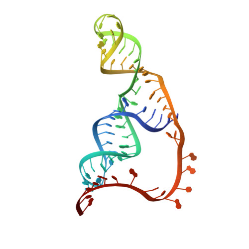
2N8V - PubMed Abstract:
The dicistrovirus intergenic region internal ribosome entry site (IRES) adopts a triple-pseudoknotted RNA structure and occupies the core ribosomal E, P, and A sites to directly recruit the ribosome and initiate translation at a non-AUG codon. A subset of dicistrovirus IRESs directs translation in the 0 and +1 frames to produce the viral structural proteins and a +1 overlapping open reading frame called ORFx, respectively. Here we show that specific mutations of two unpaired adenosines located at the core of the three-helical junction of the honey bee dicistrovirus Israeli acute paralysis virus (IAPV) IRES PKI domain can uncouple 0 and +1 frame translation, suggesting that the structure adopts distinct conformations that contribute to 0 or +1 frame translation. Using a reconstituted translation system, we show that ribosomes assembled on mutant IRESs that direct exclusive 0 or +1 frame translation lack reading frame fidelity. Finally, a nuclear magnetic resonance/small-angle X-ray scattering hybrid approach reveals that the PKI domain of the IAPV IRES adopts an RNA structure that resembles a complete tRNA. The tRNA shape-mimicry enables the viral IRES to gain access to the ribosome tRNA-binding sites and form intermolecular contacts with the ribosome that are necessary for initiating IRES translation in a specific reading frame.
- Department of Biochemistry and Molecular Biology, University of British Columbia, Vancouver, BC, Canada V6T 1Z3.
Organizational Affiliation: