2MYS | pdb_00002mys

MYOSIN SUBFRAGMENT-1, ALPHA CARBON COORDINATES ONLY FOR THE TWO LIGHT CHAINS


Experimental Data Snapshot

  • Method: X-RAY DIFFRACTION
  • Resolution: 2.80 Å
  • R-Value Work: 
    0.223 (Depositor) 

wwPDB Validation 3D Report Full Report

Validation slider image for 2MYS

This is version 1.3 of the entry. See complete history

Literature

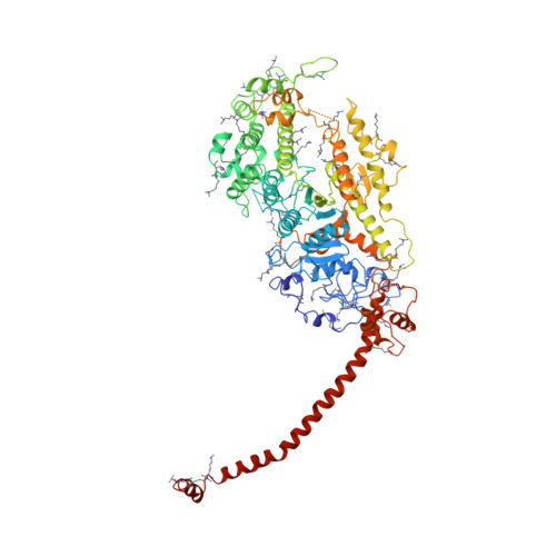
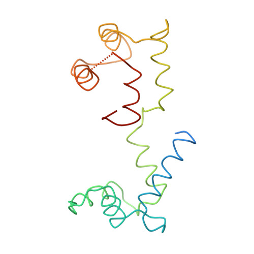
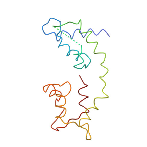
Three-dimensional structure of myosin subfragment-1: a molecular motor.

Rayment, I.Rypniewski, W.R.Schmidt-Base, K.Smith, R.Tomchick, D.R.Benning, M.M.Winkelmann, D.A.Wesenberg, G.Holden, H.M.

(1993) Science 261: 50-58

  • DOI: https://doi.org/10.1126/science.8316857
  • Primary Citation Related Structures: 
    2MYS

  • PubMed Abstract: 

    Directed movement is a characteristic of many living organisms and occurs as a result of the transformation of chemical energy into mechanical energy. Myosin is one of three families of molecular motors that are responsible for cellular motility. The three-dimensional structure of the head portion of myosin, or subfragment-1, which contains both the actin and nucleotide binding sites, is described. This structure of a molecular motor was determined by single crystal x-ray diffraction. The data provide a structural framework for understanding the molecular basis of motility.


  • Organizational Affiliation
    • Department of Biochemistry, University of Wisconsin, Madison 53705.

Macromolecule Content 

  • Total Structure Weight: 131.67 kDa 
  • Atom Count: 6,788 
  • Modeled Residue Count: 1,072 
  • Deposited Residue Count: 1,158 
  • Unique protein chains: 3

Macromolecules

Find similar proteins by:|  3D Structure
Entity ID: 1
MoleculeChains  Sequence LengthOrganismDetailsImage
MYOSIN843Gallus gallusMutation(s): 44 
UniProt
Find proteins for P13538 (Gallus gallus)
Explore P13538 
Go to UniProtKB:  P13538
Entity Groups
Sequence Clusters30% Identity50% Identity70% Identity90% Identity95% Identity100% Identity
UniProt GroupP13538
Sequence Annotations
Expand
Reference Sequence
Find similar proteins by:|  3D Structure
Entity ID: 2
MoleculeChains  Sequence LengthOrganismDetailsImage
MYOSIN166Gallus gallusMutation(s): 0 
UniProt
Find proteins for P02609 (Gallus gallus)
Explore P02609 
Go to UniProtKB:  P02609
Entity Groups
Sequence Clusters30% Identity50% Identity70% Identity90% Identity95% Identity100% Identity
UniProt GroupP02609
Sequence Annotations
Expand
Reference Sequence
Find similar proteins by:|  3D Structure
Entity ID: 3
MoleculeChains  Sequence LengthOrganismDetailsImage
MYOSIN149Gallus gallusMutation(s): 0 
UniProt
Find proteins for P02604 (Gallus gallus)
Explore P02604 
Go to UniProtKB:  P02604
Entity Groups
Sequence Clusters30% Identity50% Identity70% Identity90% Identity95% Identity100% Identity
UniProt GroupP02604
Sequence Annotations
Expand
Reference Sequence

Small Molecules

Ligands 2 Unique
IDChains Name / Formula / InChI Key2D Diagram3D Interactions
SO4

Query on SO4


Download:Ideal Coordinates CCD File
D [auth A]SULFATE ION
O4 S
QAOWNCQODCNURD-UHFFFAOYSA-L
MG

Query on MG


Download:Ideal Coordinates CCD File
E [auth B]MAGNESIUM ION
Mg
JLVVSXFLKOJNIY-UHFFFAOYSA-N
Modified Residues  1 Unique
IDChains TypeFormula2D DiagramParent
MLY
Query on MLY
A
L-PEPTIDE LINKINGC8 H18 N2 O2LYS

Experimental Data & Validation

Experimental Data

  • Method: X-RAY DIFFRACTION
  • Resolution: 2.80 Å
  • R-Value Work:  0.223 (Depositor) 
Space Group: C 2 2 21
Unit Cell:
Length ( Å )Angle ( ˚ )
a = 98.4α = 90
b = 124.2β = 90
c = 274.9γ = 90
Software Package:
Software NamePurpose
TNTrefinement
XDSdata reduction
ROSSMANNdata reduction
CCP4data scaling

Structure Validation

View Full Validation Report


Entry History 

Deposition Data

Revision History  (Full details and data files)

  • Version 1.0: 1997-01-11
    Type: Initial release
  • Version 1.1: 2008-03-24
    Changes: Version format compliance
  • Version 1.2: 2011-07-13
    Changes: Version format compliance
  • Version 1.3: 2024-06-05
    Changes: Data collection, Database references, Derived calculations, Other