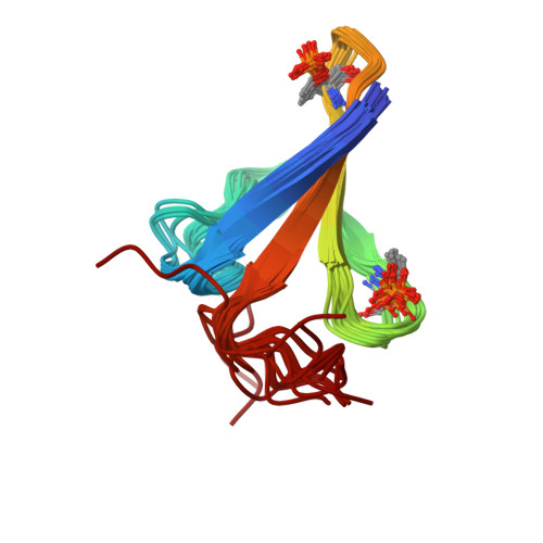
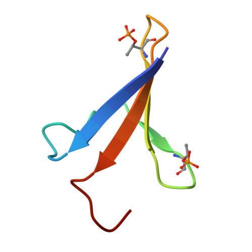
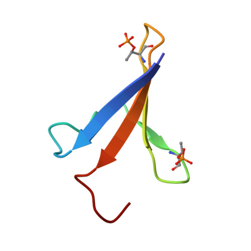
Folding of an intrinsically disordered protein by phosphorylation as a regulatory switch.
Bah, A., Vernon, R.M., Siddiqui, Z., Krzeminski, M., Muhandiram, R., Zhao, C., Sonenberg, N., Kay, L.E., Forman-Kay, J.D.(2015) Nature 519: 106-109
- PubMed: 25533957
- DOI: https://doi.org/10.1038/nature13999
- Primary Citation of Related Structures:
2MX4 - PubMed Abstract:
Intrinsically disordered proteins play important roles in cell signalling, transcription, translation and cell cycle regulation. Although they lack stable tertiary structure, many intrinsically disordered proteins undergo disorder-to-order transitions upon binding to partners. Similarly, several folded proteins use regulated order-to-disorder transitions to mediate biological function. In principle, the function of intrinsically disordered proteins may be controlled by post-translational modifications that lead to structural changes such as folding, although this has not been observed. Here we show that multisite phosphorylation induces folding of the intrinsically disordered 4E-BP2, the major neural isoform of the family of three mammalian proteins that bind eIF4E and suppress cap-dependent translation initiation. In its non-phosphorylated state, 4E-BP2 interacts tightly with eIF4E using both a canonical YXXXXLΦ motif (starting at Y54) that undergoes a disorder-to-helix transition upon binding and a dynamic secondary binding site. We demonstrate that phosphorylation at T37 and T46 induces folding of residues P18-R62 of 4E-BP2 into a four-stranded β-domain that sequesters the helical YXXXXLΦ motif into a partly buried β-strand, blocking its accessibility to eIF4E. The folded state of pT37pT46 4E-BP2 is weakly stable, decreasing affinity by 100-fold and leading to an order-to-disorder transition upon binding to eIF4E, whereas fully phosphorylated 4E-BP2 is more stable, decreasing affinity by a factor of approximately 4,000. These results highlight stabilization of a phosphorylation-induced fold as the essential mechanism for phospho-regulation of the 4E-BP:eIF4E interaction and exemplify a new mode of biological regulation mediated by intrinsically disordered proteins.
Organizational Affiliation:
1] Molecular Structure and Function Program, Hospital for Sick Children, Toronto, Ontario M5G 0A4, Canada [2] Department of Biochemistry, University of Toronto, Toronto, Ontario M5S 1A8, Canada.


