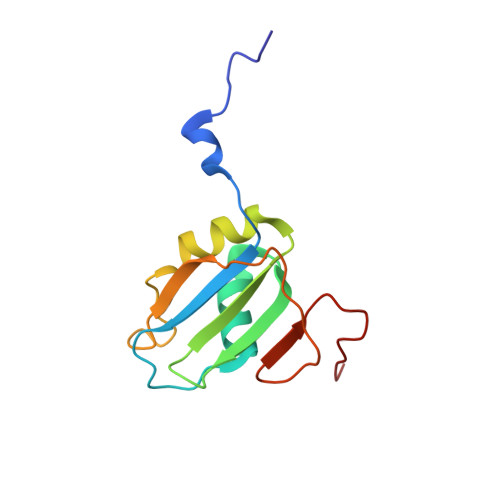
The Signature of the Five-Stranded vRRM Fold Defined by Functional, Structural and Computational Analysis of the hnRNP L Protein.
Blatter, M., Dunin-Horkawicz, S., Grishina, I., Maris, C., Thore, S., Maier, T., Bindereif, A., Bujnicki, J.M., Allain, F.H.(2015) J Mol Biology 427: 3001-3022
- PubMed: 26051023 Search on PubMed
- DOI: https://doi.org/10.1016/j.jmb.2015.05.020
- Primary Citation Related Structures:
2MQL, 2MQM, 2MQN, 4QPT - PubMed Abstract:
The RNA recognition motif (RRM) is the far most abundant RNA binding domain. In addition to the typical β1α1β2β3α2β4 fold, various sub-structural elements have been described and reportedly contribute to the high functional versatility of RRMs. The heterogeneous nuclear ribonucleoprotein L (hnRNP L) is a highly abundant protein of 64 kDa comprising four RRM domains. Involved in many aspects of RNA metabolism, hnRNP L specifically binds to RNAs containing CA repeats or CA-rich clusters. However, a comprehensive structural description of hnRNP L including its sub-structural elements is missing. Here, we present the structural characterization of the RRM domains of hnRNP L and demonstrate their function in repressing exon 4 of SLC2A2. By comparison of the sub-structural elements between the two highly similar paralog families of hnRNP L and PTB, we defined signatures underlying interacting C-terminal coils (ICCs), the RRM34 domain interaction and RRMs with a C-terminal fifth β-strand, a variation we denoted vRRMs. Furthermore, computational analysis revealed new putative ICC-containing RRM families and allowed us to propose an evolutionary scenario explaining the origins of the ICC and fifth β-strand sub-structural extensions. Our studies provide insights of domain requirements in alternative splicing mediated by hnRNP L and molecular descriptions for the sub-structural elements. In addition, the analysis presented may help to classify other abundant RRM extensions and to predict structure-function relationships.
- Institute of Molecular Biology and Biophysics, ETH Zurich, 8093 Zurich, Switzerland.
Organizational Affiliation: