2MGV | pdb_00002mgv

NMR structure of PASTA domain of PonA2 from Mycobacterium tuberculosis


Experimental Data Snapshot

  • Method: SOLUTION NMR
  • Conformers Calculated: 150 
  • Conformers Submitted: 10 
  • Selection Criteria: structures with the least restraint violations 

wwPDB Validation 3D Report Full Report

Validation slider image for 2MGV

This is version 1.3 of the entry. See complete history

Literature

Structural and binding properties of the PASTA domain of PonA2, a key penicillin binding protein from Mycobacterium tuberculosis.

Calvanese, L.Falcigno, L.Maglione, C.Marasco, D.Ruggiero, A.Squeglia, F.Berisio, R.D'Auria, G.

(2014) Biopolymers 101: 712-719

  • DOI: https://doi.org/10.1002/bip.22447
  • Primary Citation Related Structures: 
    2MGV

  • PubMed Abstract: 

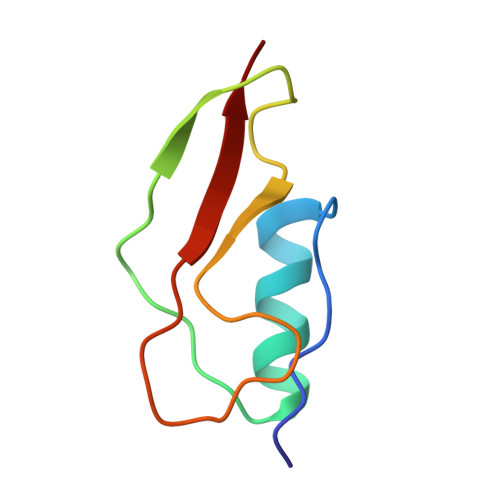
    PonA2 is one of the two class A penicillin binding proteins of Mycobacterium tuberculosis, the etiologic agent of tuberculosis. It plays a complex role in mycobacterial physiology and is spotted as a promising target for inhibitors. PonA2 is involved in adaptation of M. tuberculosis to dormancy, an ability which has been attributed to the presence in its sequence of a C-terminal PASTA domain. Since PASTA modules are typically considered as β-lactam antibiotic binding domains, we determined the solution structure of the PASTA domain from PonA2 and analyzed its binding properties versus a plethora of potential binders, including the β-lactam antibiotics, two typical muropeptide mimics, and polymeric peptidoglycan. We show that, despite a high structural similarity with other PASTA domains, the PASTA domain of PonA2 displays different binding properties, as it is not able to bind muropeptides, or β-lactams, or polymeric peptidoglycan. These results indicate that the role of PASTA domains cannot be generalized, as their specific binding properties strongly depend on surface residues, which are widely variable.


  • Organizational Affiliation
    • CIRPeB, University of Naples Federico II, Naples, Italy.

Macromolecule Content 

  • Total Structure Weight: 6.58 kDa 
  • Atom Count: 461 
  • Modeled Residue Count: 65 
  • Deposited Residue Count: 65 
  • Unique protein chains: 1

Macromolecules

Find similar proteins by:|  3D Structure
Entity ID: 1
MoleculeChains  Sequence LengthOrganismDetailsImage
Bifunctional membrane-associated penicillin-binding protein 1A/1B ponA265Mycobacterium tuberculosis H37RvMutation(s): 0 
Gene Names: RVBD_3682
EC: 2.4.99.28 (UniProt), 3.4.16.4 (UniProt)
UniProt
Find proteins for I6YGX2 (Mycobacterium tuberculosis (strain ATCC 25618 / H37Rv))
Explore I6YGX2 
Go to UniProtKB:  I6YGX2
Entity Groups
Sequence Clusters30% Identity50% Identity70% Identity90% Identity95% Identity100% Identity
UniProt GroupI6YGX2
Sequence Annotations
Expand
Reference Sequence

Experimental Data & Validation

Experimental Data

  • Method: SOLUTION NMR
  • Conformers Calculated: 150 
  • Conformers Submitted: 10 
  • Selection Criteria: structures with the least restraint violations 

Structure Validation

View Full Validation Report


Entry History 

Deposition Data

Revision History  (Full details and data files)

  • Version 1.0: 2013-12-25
    Type: Initial release
  • Version 1.1: 2014-05-07
    Changes: Database references
  • Version 1.2: 2023-06-14
    Changes: Data collection, Database references, Other
  • Version 1.3: 2024-05-15
    Changes: Data collection, Database references