2MFC | pdb_00002mfc

Csr/Rsm protein-RNA recognition - A molecular affinity ruler: RsmZ(SL1)/RsmE(dimer) 2:1 complex


Experimental Data Snapshot

  • Method: SOLUTION NMR
  • Conformers Calculated: 999 
  • Conformers Submitted: 20 
  • Selection Criteria: structures with the lowest energy 

wwPDB Validation 3D Report Full Report

Validation slider image for 2MFC

This is version 1.3 of the entry. See complete history

Literature

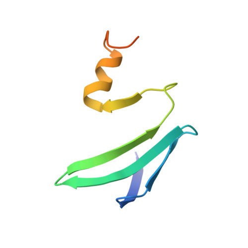
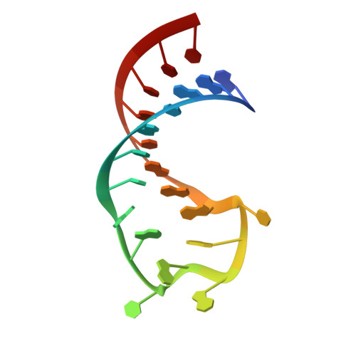
Molecular basis for the wide range of affinity found in Csr/Rsm protein-RNA recognition.

Duss, O.Michel, E.Diarra Dit Konte, N.Schubert, M.Allain, F.H.

(2014) Nucleic Acids Res 42: 5332-5346

  • DOI: https://doi.org/10.1093/nar/gku141
  • Primary Citation Related Structures: 
    2MFC, 2MFE, 2MFF, 2MFG, 2MFH

  • PubMed Abstract: 

    The carbon storage regulator/regulator of secondary metabolism (Csr/Rsm) type of small non-coding RNAs (sRNAs) is widespread throughout bacteria and acts by sequestering the global translation repressor protein CsrA/RsmE from the ribosome binding site of a subset of mRNAs. Although we have previously described the molecular basis of a high affinity RNA target bound to RsmE, it remains unknown how other lower affinity targets are recognized by the same protein. Here, we have determined the nuclear magnetic resonance solution structures of five separate GGA binding motifs of the sRNA RsmZ of Pseudomonas fluorescens in complex with RsmE. The structures explain how the variation of sequence and structural context of the GGA binding motifs modulate the binding affinity for RsmE by five orders of magnitude (∼10 nM to ∼3 mM, Kd). Furthermore, we see that conformational adaptation of protein side-chains and RNA enable recognition of different RNA sequences by the same protein contributing to binding affinity without conferring specificity. Overall, our findings illustrate how the variability in the Csr/Rsm protein-RNA recognition allows a fine-tuning of the competition between mRNAs and sRNAs for the CsrA/RsmE protein.


  • Organizational Affiliation
    • Institute of Molecular Biology and Biophysics, ETH Zürich, 8093 Zürich, Switzerland.

Macromolecule Content 

  • Total Structure Weight: 29.92 kDa 
  • Atom Count: 1,838 
  • Modeled Residue Count: 162 
  • Deposited Residue Count: 184 
  • Unique protein chains: 1
  • Unique nucleic acid chains: 1

Macromolecules


Find similar proteins by:|  3D Structure
Entity ID: 1
MoleculeChains  Sequence LengthOrganismDetailsImage
Carbon storage regulator homolog
A, C
70Pseudomonas fluorescensMutation(s): 0 
Gene Names: rsmEcsrA
UniProt
Find proteins for P0DPC3 (Pseudomonas protegens (strain DSM 19095 / LMG 27888 / CFBP 6595 / CHA0))
Explore P0DPC3 
Go to UniProtKB:  P0DPC3
Entity Groups
Sequence Clusters30% Identity50% Identity70% Identity90% Identity95% Identity100% Identity
UniProt GroupP0DPC3
Sequence Annotations
Expand
Reference Sequence
Find similar nucleic acids by:  Sequence
Entity ID: 2
MoleculeChains LengthOrganismImage
SL1(RsmZ) RNA
B, D
22N/A
Sequence Annotations
Expand
Reference Sequence

Experimental Data & Validation

Experimental Data

  • Method: SOLUTION NMR
  • Conformers Calculated: 999 
  • Conformers Submitted: 20 
  • Selection Criteria: structures with the lowest energy 

Structure Validation

View Full Validation Report


Entry History 

Deposition Data

Revision History  (Full details and data files)

  • Version 1.0: 2014-02-26
    Type: Initial release
  • Version 1.1: 2014-03-26
    Changes: Database references
  • Version 1.2: 2014-05-14
    Changes: Database references
  • Version 1.3: 2024-05-01
    Changes: Data collection, Database references