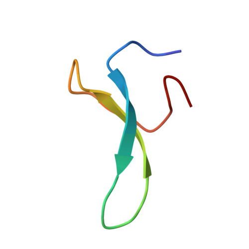
2M9J

NMR solution structure of Pin1 WW domain mutant 6-1g


Experimental Data Snapshot

  • Method: SOLUTION NMR
  • Conformers Calculated: 200 
  • Conformers Submitted: 17 
  • Selection Criteria: structures with the lowest energy 

wwPDB Validation   3D Report Full Report


This is version 1.4 of the entry. See complete history


Literature

Structural and energetic basis of carbohydrate-aromatic packing interactions in proteins.

Chen, W.Enck, S.Price, J.L.Powers, D.L.Powers, E.T.Wong, C.H.Dyson, H.J.Kelly, J.W.

(2013) J Am Chem Soc 135: 9877-9884

  • DOI: https://doi.org/10.1021/ja4040472
  • Primary Citation of Related Structures:  
    2M9E, 2M9F, 2M9I, 2M9J

  • PubMed Abstract: 

    Carbohydrate-aromatic interactions mediate many biological processes. However, the structure-energy relationships underpinning direct carbohydrate-aromatic packing interactions in aqueous solution have been difficult to assess experimentally and remain elusive. Here, we determine the structures and folding energetics of chemically synthesized glycoproteins to quantify the contributions of the hydrophobic effect and CH-π interactions to carbohydrate-aromatic packing interactions in proteins. We find that the hydrophobic effect contributes significantly to protein-carbohydrate interactions. Interactions between carbohydrates and aromatic amino acid side chains, however, are supplemented by CH-π interactions. The strengths of experimentally determined carbohydrate CH-π interactions do not correlate with the electrostatic properties of the involved aromatic residues, suggesting that the electrostatic component of CH-π interactions in aqueous solution is small. Thus, tight binding of carbohydrates and aromatic residues is driven by the hydrophobic effect and CH-π interactions featuring a dominating dispersive component.


  • Organizational Affiliation

    Department of Molecular and Experimental Medicine, The Scripps Research Institute, La Jolla, California 92037, USA.


Macromolecules
Find similar proteins by:  (by identity cutoff)  |  3D Structure
Entity ID: 1
MoleculeChains Sequence LengthOrganismDetailsImage
Peptidyl-prolyl cis-trans isomerase NIMA-interacting 134Homo sapiensMutation(s): 0 
EC: 5.2.1.8
UniProt & NIH Common Fund Data Resources
Find proteins for Q13526 (Homo sapiens)
Explore Q13526 
Go to UniProtKB:  Q13526
PHAROS:  Q13526
GTEx:  ENSG00000127445 
Entity Groups  
Sequence Clusters30% Identity50% Identity70% Identity90% Identity95% Identity100% Identity
UniProt GroupQ13526
Glycosylation
Glycosylation Sites: 1Go to GlyGen: Q13526-1
Sequence Annotations
Expand
  • Reference Sequence
Small Molecules
Ligands 1 Unique
IDChains Name / Formula / InChI Key2D Diagram3D Interactions
NAG
Query on NAG

Download Ideal Coordinates CCD File 
B [auth A]2-acetamido-2-deoxy-beta-D-glucopyranose
C8 H15 N O6
OVRNDRQMDRJTHS-FMDGEEDCSA-N
Experimental Data & Validation

Experimental Data

  • Method: SOLUTION NMR
  • Conformers Calculated: 200 
  • Conformers Submitted: 17 
  • Selection Criteria: structures with the lowest energy 

Structure Validation

View Full Validation Report


Entry History 

Deposition Data

Revision History  (Full details and data files)

  • Version 1.0: 2013-06-26
    Type: Initial release
  • Version 1.1: 2013-07-03
    Changes: Database references
  • Version 1.2: 2013-07-17
    Changes: Database references
  • Version 1.3: 2020-07-29
    Type: Remediation
    Reason: Carbohydrate remediation
    Changes: Data collection, Derived calculations, Structure summary
  • Version 1.4: 2024-11-27
    Changes: Data collection, Database references, Structure summary