2M66 | pdb_00002m66

Endoplasmic reticulum protein 29 (ERp29) C-terminal domain: 3D Protein Fold Determination from Backbone Amide Pseudocontact Shifts Generated by Lanthanide Tags at Multiple Sites


Experimental Data Snapshot

  • Method: SOLUTION NMR
  • Conformers Calculated: 100000 
  • Conformers Submitted: 11 
  • Selection Criteria: target function 

wwPDB Validation 3D Report Full Report

Validation slider image for 2M66

This is version 1.1 of the entry. See complete history

Literature

Three-dimensional protein fold determination from backbone amide pseudocontact shifts generated by lanthanide tags at multiple sites

Yagi, H.Pilla, K.B.Maleckis, A.Graham, B.Huber, T.Otting, G.

(2013) Structure 21: 883-890

  • DOI: https://doi.org/10.1016/j.str.2013.04.001
  • Primary Citation Related Structures: 
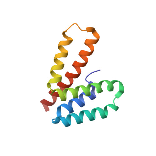
    2M66

  • PubMed Abstract: 

    Site-specific attachment of paramagnetic lanthanide ions to a protein generates pseudocontact shifts (PCS) in the nuclear magnetic resonance (NMR) spectra of the protein that are easily measured as changes in chemical shifts. By labeling the protein with lanthanide tags at four different sites, PCSs are observed for most amide protons and accurate information is obtained about their coordinates in three-dimensional space. The approach is demonstrated with the chaperone ERp29, for which large differences have been reported between X-ray and NMR structures of the C-terminal domain, ERp29-C. The results unambiguously show that the structure of rat ERp29-C in solution is similar to the crystal structure of human ERp29-C. PCSs of backbone amides were the only structural restraints required. Because these can be measured for more dilute protein solutions than other NMR restraints, the approach greatly widens the range of proteins amenable to structural studies in solution.


  • Organizational Affiliation
    • Research School of Chemistry, Australian National University, Canberra, ACT 0200, Australia.

Macromolecule Content 

  • Total Structure Weight: 11.79 kDa 
  • Atom Count: 826 
  • Modeled Residue Count: 106 
  • Deposited Residue Count: 106 
  • Unique protein chains: 1

Macromolecules

Find similar proteins by:|  3D Structure
Entity ID: 1
MoleculeChains  Sequence LengthOrganismDetailsImage
Endoplasmic reticulum resident protein 29106Rattus norvegicusMutation(s): 0 
Gene Names: Erp29
UniProt
Find proteins for P52555 (Rattus norvegicus)
Explore P52555 
Go to UniProtKB:  P52555
Entity Groups
Sequence Clusters30% Identity50% Identity70% Identity90% Identity95% Identity100% Identity
UniProt GroupP52555
Sequence Annotations
Expand
Reference Sequence

Experimental Data & Validation

Experimental Data

  • Method: SOLUTION NMR
  • Conformers Calculated: 100000 
  • Conformers Submitted: 11 
  • Selection Criteria: target function 

Structure Validation

View Full Validation Report


Entry History 

Deposition Data

Revision History  (Full details and data files)

  • Version 1.0: 2013-07-10
    Type: Initial release
  • Version 1.1: 2024-05-01
    Changes: Data collection, Database references