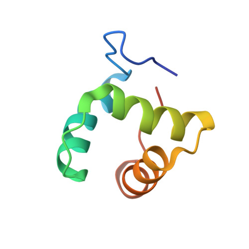
Structures of the Compact Helical Core Domains of Feline Calicivirus and Murine Norovirus VPg Proteins.
Leen, E.N., Kwok, K.Y., Birtley, J.R., Simpson, P.J., Subba-Reddy, C.V., Chaudhry, Y., Sosnovtsev, S.V., Green, K.Y., Prater, S.N., Tong, M., Young, J.C., Chung, L.M., Marchant, J., Roberts, L.O., Kao, C.C., Matthews, S., Goodfellow, I.G., Curry, S.(2013) J Virol 87: 5318-5330
- PubMed: 23487472
- DOI: https://doi.org/10.1128/JVI.03151-12
- Primary Citation of Related Structures:
2M4G, 2M4H - PubMed Abstract:
We report the solution structures of the VPg proteins from feline calicivirus (FCV) and murine norovirus (MNV), which have been determined by nuclear magnetic resonance spectroscopy. In both cases, the core of the protein adopts a compact helical structure flanked by flexible N and C termini. Remarkably, while the core of FCV VPg contains a well-defined three-helix bundle, the MNV VPg core has just the first two of these secondary structure elements. In both cases, the VPg cores are stabilized by networks of hydrophobic and salt bridge interactions. The Tyr residue in VPg that is nucleotidylated by the viral NS7 polymerase (Y24 in FCV, Y26 in MNV) occurs in a conserved position within the first helix of the core. Intriguingly, given its structure, VPg would appear to be unable to bind to the viral polymerase so as to place this Tyr in the active site without a major conformation change to VPg or the polymerase. However, mutations that destabilized the VPg core either had no effect on or reduced both the ability of the protein to be nucleotidylated and virus infectivity and did not reveal a clear structure-activity relationship. The precise role of the calicivirus VPg core in virus replication remains to be determined, but knowledge of its structure will facilitate future investigations.
Organizational Affiliation:
Division of Cell and Molecular Biology, Department of Life Sciences, Imperial College London, London, United Kingdom.


