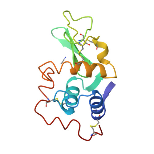
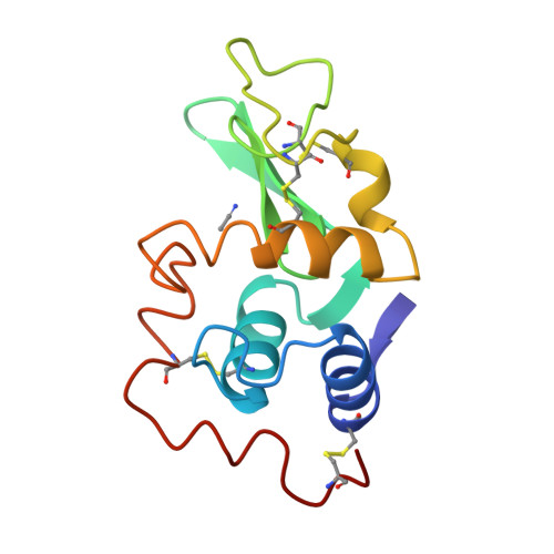
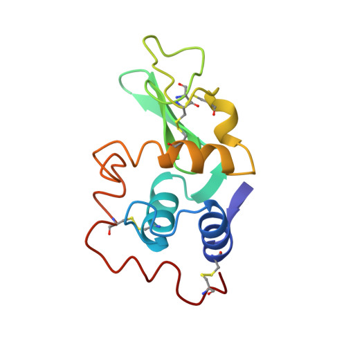
X-ray studies on cross-linked lysozyme crystals in acetonitrile-water mixture.
Wang, Z., Zhu, G., Huang, Q., Qian, M., Shao, M., Jia, Y., Tang, Y.(1998) Biochim Biophys Acta 1384: 335-344
- PubMed: 9659395
- DOI: https://doi.org/10.1016/s0167-4838(98)00027-2
- Primary Citation of Related Structures:
1LYO, 2LYO, 3LYO, 4LYO - PubMed Abstract:
Tetragonal crystals of hen egg white lysozyme were cross-linked and subjected to X-ray diffraction study in acetonitrile-water media with different acetonitrile concentrations. Crystals in neat acetonitrile did not scatter X-ray well. Structures of crystals in neat water, in 90% and 95% acetonitrile, and crystal back-soaked from acetonitrile to water, were determined to about 2 A resolution. For crystals in both 90% acetonitrile, and crystal back-soaked from acetonitrile to water, were determined to about 2 A resolution. For crystals in both 90% and 95% acetonitrile, only one protein-bond acetonitrile molecule is found in the active site cleft, and its location and binding-protein mode is similar to the C subunit of polysaccharide. The alteration in conformation and hydrogen-bond pattern involving water as solvent causes the reduction of the protein's flexibility in organic media. The back-soaked crystal regained its ordinary three-dimensional structure in water.
Organizational Affiliation:
Department of Chemistry, Peking University, Beijing, China.


