Conformational analysis of StrH, the surface-attached exo-beta-D-N-acetylglucosaminidase from Streptococcus pneumoniae.
Pluvinage, B., Chitayat, S., Ficko-Blean, E., Abbott, D.W., Kunjachen, J.M., Grondin, J., Spencer, H.L., Smith, S.P., Boraston, A.B.(2013) J Mol Biology 425: 334-349
- PubMed: 23154168 Search on PubMed
- DOI: https://doi.org/10.1016/j.jmb.2012.11.005
- Primary Citation Related Structures:
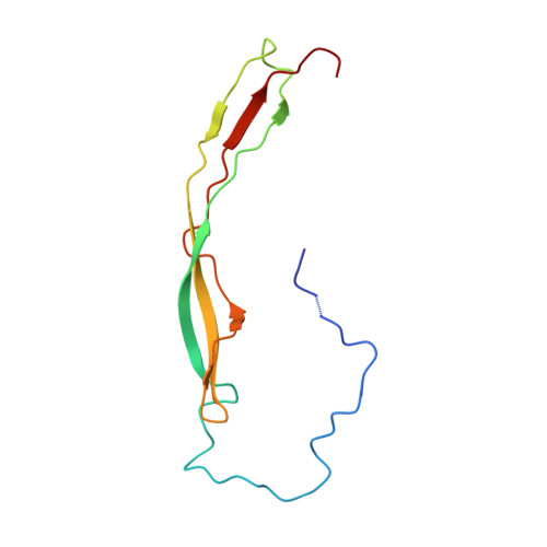
2LTJ - PubMed Abstract:
Streptococcus pneumoniae is a serious human pathogen that presents on its surface numerous proteins involved in the host-bacterium interaction. The carbohydrate-active enzymes are particularly well represented among these surface proteins, and many of these are known virulence factors, highlighting the importance of carbohydrate processing by this pathogen. StrH is a surface-attached exo-β-D-N-acetylglucosaminidase that cooperates with the sialidase NanA and the β-galactosidase BgaA to sequentially degrade the nonreducing terminal arms of complex N-linked glycans. This enzyme is a large multi-modular protein that is notable for its tandem N-terminal family GH20 catalytic modules, whose individual X-ray crystal structures were recently reported. StrH also contains C-terminal tandem G5 modules, which are uncharacterized. Here, we report the NMR-determined solution structure of the first G5 module in the tandem, G5-1, which along with the X-ray crystal structures of the GH20 modules was used in conjunction with small-angle X-ray scattering to construct a pseudo-atomic model of full-length StrH. The results reveal a model in which StrH adopts an elongated conformation that may project the catalytic modules away from the surface of the bacterium to a distance of up to ~250 Å.
- Biochemistry and Microbiology, University of Victoria, Victoria, BC, Canada V8W 3P6.
Organizational Affiliation: