Refinement of a molecular model for lamprey hemoglobin from Petromyzon marinus.
Honzatko, R.B., Hendrickson, W.A., Love, W.E.(1985) J Mol Biology 184: 147-164
- PubMed: 4032476 Search on PubMed
- DOI: https://doi.org/10.1016/0022-2836(85)90049-x
- Primary Citation Related Structures:
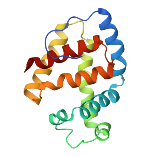
2LHB - PubMed Abstract:
A molecular model for the protein and ambient solvent of the complex of cyanide with methemoglobin V from the sea lamprey Petromyzon marinus yields an R-factor of 0.142 against X-ray diffraction data to 2.0 A resolution. The root-mean-square discrepancies from ideal bond length and angle are, respectively, 0.014 A and 1.5 degrees. Atoms that belong to planar groups deviate by 0.012 A from planes determined by a least-squares procedure. The average standard deviation for chiral volumes, peptide torsion angle and torsion angles of side-chains are 0.150 A3, 2.0 degrees and 19.4 degrees, respectively. The root-mean-square variation in the thermal parameters of bonded atoms of the polypeptide backbone is 1.21 A2; the variation in thermal parameters for side-chain atoms is 2.13 A2. The model includes multiple conformations for 11 side-chains of the 149 amino acid residues of the protein. We identify 231 locations as sites of water molecules in full or partial occupancy. The sum of occupancy factors for these sites is approximately 154, representing 28% of the 550 molecules of water within the crystallographic asymmetric unit. The environment of the heme in the cyanide complex of lamprey methemoglobin resembles the deoxy state of the mammalian tetramer. In particular, the bond between atom NE2 of the proximal histidine and the Fe lies 5.1 degrees from the normal of the heme plane. In deoxy- and carbonmonoxyhemoglobins, the deviations from the normal to the heme plane are 7 to 8 degrees and 1 degree, respectively. Furthermore, the inequality in the distance of atom CD2 of the proximal histidine from the pyrrole nitrogen of ring-C of the heme (distance = 3.29 A) and CE1 from the pyrrole nitrogen of ring-A (distance = 3.06 A) is characteristic of deoxyhemoglobin, not carbonmonoxyhemoglobin, where these distances are equal. Finally, a hydrogen bond exists between carbonyl 111 and the hydroxyl of tyrosine 149. The corresponding hydrogen link in the mammalian tetramer is central to the T to R state transition and is present in deoxyhemoglobin but absent in carbonmonoxyhemoglobin. We suggest that the low affinity of oxygen for lamprey hemoglobin may be a consequence of these T-state geometries.