2LFO | pdb_00002lfo

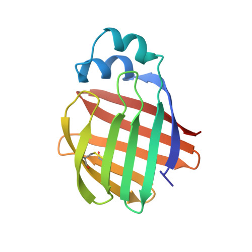
NMR structure of cl-BABP/SS complexed with glycochenodeoxycholic and glycocholic acids


Experimental Data Snapshot

  • Method: SOLUTION NMR
  • Conformers Calculated: 50 
  • Conformers Submitted: 20 
  • Selection Criteria: best HADDOCK score 

wwPDB Validation 3D Report Full Report

Validation slider image for 2LFO

This is version 1.2 of the entry. See complete history. 

Literature

A disulfide bridge allows for site-selective binding in liver bile acid binding protein thereby stabilising the orientation of key amino acid side chains.

Tomaselli, S., Assfalg, M., Pagano, K., Cogliati, C., Zanzoni, S., Molinari, H., Ragona, L.

(2012) Chemistry 18: 2857-2866

  • DOI: https://doi.org/10.1002/chem.201102203
  • Primary Citation Related Structures: 
    2LFO

  • PubMed Abstract: 

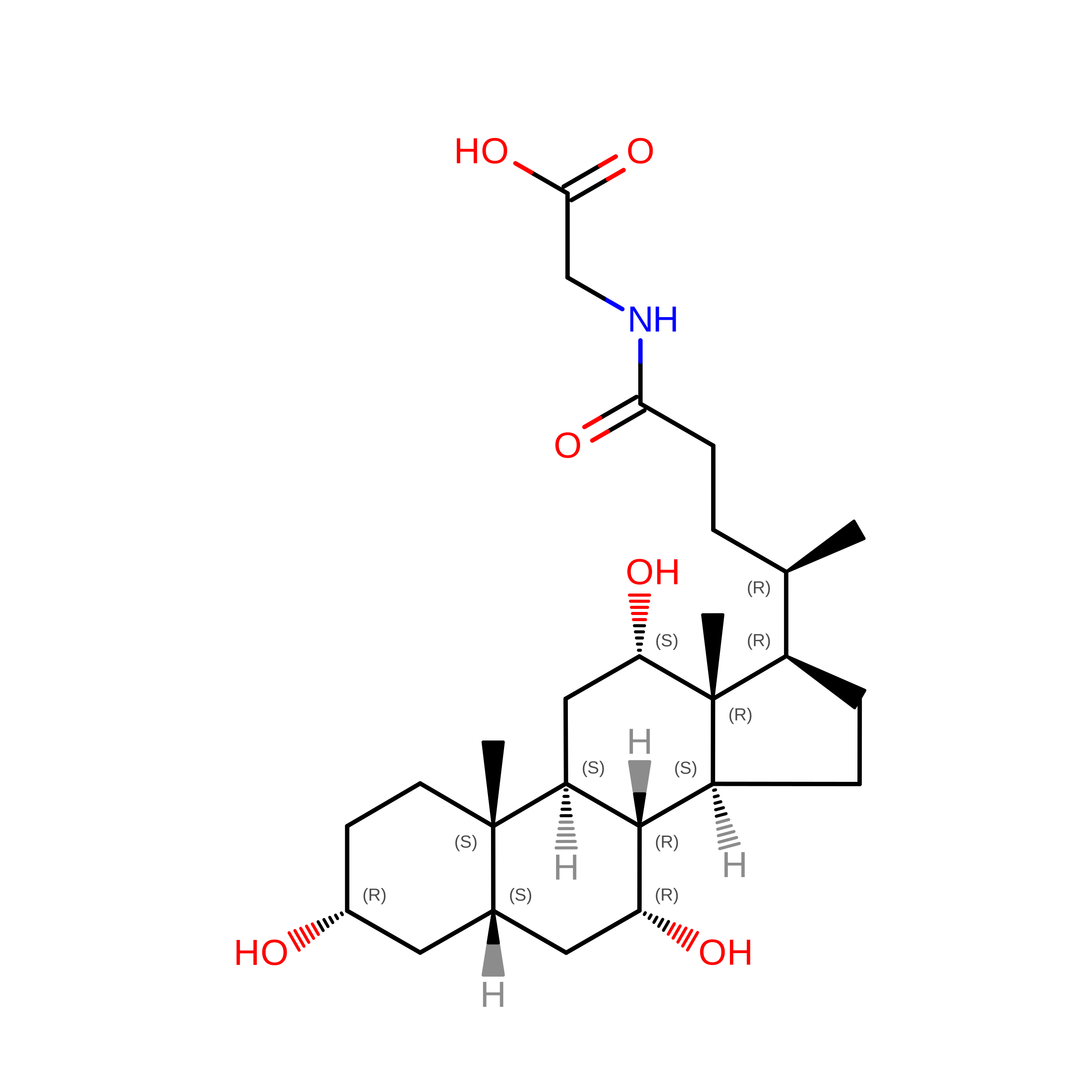
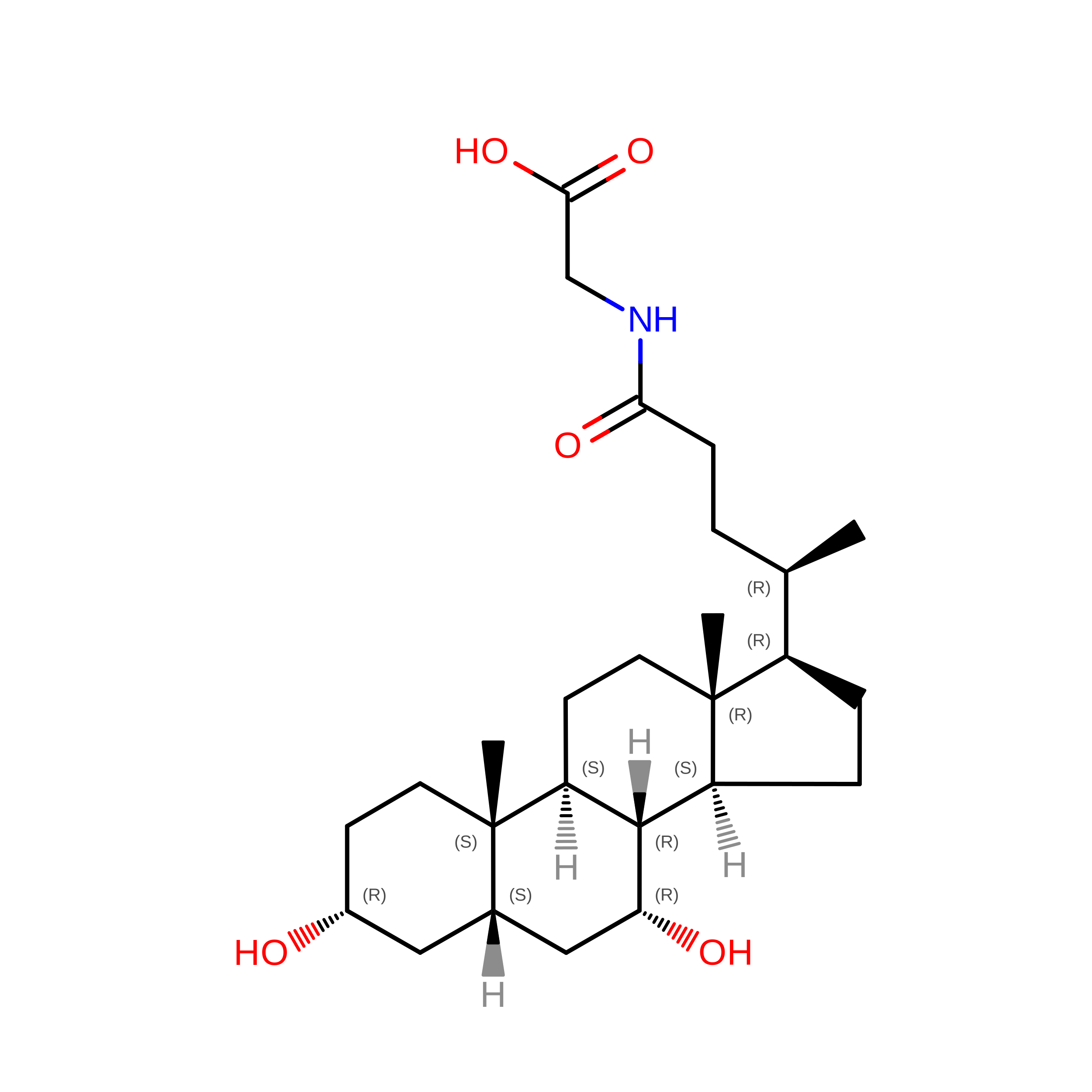
    The presence of a disulfide bridge in liver bile acid binding protein (L-BABP/S-S) allows for site-selective binding of two bile acids, glycochenodeoxycholic (GCDA) and glycocholic acid (GCA), differing only in the presence of a hydroxyl group. The protein form devoid of the disulfide bridge (L-BABP) binds both bile salts without discriminating ability. We investigate the determinants of the molecular recognition process in the formation of the heterotypic L-BABP/S-S complex with GCDA [corrected] and GCA [corrected] located in the superficial and inner protein sites, respectively. The comparison of the NMR spectroscopy structure of heterotypic holo L-BABP/S-S, the first reported for this protein family, with that of the homotypic L-BABP complex demonstrates that the introduction of a S-S link between adjacent strands changes the conformation of three key residues, which function as hot-spot mediators of molecular discrimination. The favoured χ(1) rotameric states (t, g(+) and g(-) for E99, Q100 and E109 residues, respectively) allow the onset of an extended intramolecular hydrogen-bond network and the consequent stabilisation of the side-chain orientation of a buried histidine, which is capable of anchoring a specific ligand.


  • Organizational Affiliation: 
    • Laboratorio NMR, ISMAC-CNR, Via Bassini 15, 20133 Milano, Italy.

Macromolecule Content 

  • Total Structure Weight: 15.02 kDa 
  • Atom Count: 1,053 
  • Modeled Residue Count: 125 
  • Deposited Residue Count: 125 
  • Unique protein chains: 1

Macromolecules

Find similar proteins by:|  3D Structure
Entity ID: 1
MoleculeChains  Sequence LengthOrganismDetailsImage
Fatty acid-binding protein, liver125Gallus gallusMutation(s): 1 
Gene Names: FABP1
UniProt
Find proteins for P80226 (Gallus gallus)
Explore P80226 
Go to UniProtKB:  P80226
Entity Groups
Sequence Clusters30% Identity50% Identity70% Identity90% Identity95% Identity100% Identity
UniProt GroupP80226
Sequence Annotations
Expand
Reference Sequence

Small Molecules

Ligands 2 Unique
IDChains Name / Formula / InChI Key2D Diagram3D Interactions
GCH

Query on GCH


Download:Ideal Coordinates CCD File
B [auth A]GLYCOCHOLIC ACID
C26 H43 N O6
RFDAIACWWDREDC-FRVQLJSFSA-N
CHO

Query on CHO


Download:Ideal Coordinates CCD File
C [auth A]GLYCOCHENODEOXYCHOLIC ACID
C26 H43 N O5
GHCZAUBVMUEKKP-GYPHWSFCSA-N

Experimental Data & Validation

Experimental Data

  • Method: SOLUTION NMR
  • Conformers Calculated: 50 
  • Conformers Submitted: 20 
  • Selection Criteria: best HADDOCK score 

Structure Validation

View Full Validation Report


Entry History 

Deposition Data

Revision History  (Full details and data files)

  • Version 1.0: 2012-07-11
    Type: Initial release
  • Version 1.1: 2023-06-14
    Changes: Data collection, Database references, Derived calculations, Other
  • Version 1.2: 2024-10-30
    Changes: Data collection, Database references, Structure summary