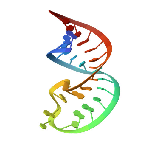
Solution Structure of the HIV-1 Exon Splicing Silencer 3.
Levengood, J.D., Rollins, C., Mishler, C.H., Johnson, C.A., Miner, G., Rajan, P., Znosko, B.M., Tolbert, B.S.(2012) J Mol Biol 415: 680-698
- PubMed: 22154809 Search on PubMed
- DOI: https://doi.org/10.1016/j.jmb.2011.11.034
- Primary Citation Related Structures:
2LDL - PubMed Abstract:
Alternative splicing of the human immunodeficiency virus type 1 (HIV-1) genomic RNA is necessary to produce the complete viral protein complement, and aberrations in the splicing pattern impair HIV-1 replication. Genome splicing in HIV-1 is tightly regulated by the dynamic assembly/disassembly of trans host factors with cis RNA control elements. The host protein, heterogeneous nuclear ribonucleoprotein (hnRNP) A1, regulates splicing at several highly conserved HIV-1 3' splice sites by binding 5'-UAG-3' elements embedded within regions containing RNA structure. The physical determinants of hnRNP A1 splice site recognition remain poorly defined in HIV-1, thus precluding a detailed understanding of the molecular basis of the splicing pattern. Here, the three-dimensional structure of the exon splicing silencer 3 (ESS3) from HIV-1 has been determined using NMR spectroscopy. ESS3 adopts a 27-nucleotide hairpin with a 10-bp A-form stem that contains a pH-sensitive A(+)C wobble pair. The seven-nucleotide hairpin loop contains the high-affinity hnRNP-A1-responsive 5'-UAGU-3' element and a proximal 5'-GAU-3' motif. The NMR structure shows that the heptaloop adopts a well-organized conformation stabilized primarily by base stacking interactions reminiscent of a U-turn. The apex of the loop is quasi-symmetric with UA dinucleotide steps from the 5'-GAU-3' and 5'-UAGU-3' motifs stacking on opposite sides of the hairpin. As a step towards understanding the binding mechanism, we performed calorimetric and NMR titrations of several hnRNP A1 subdomains into ESS3. The data show that the UP1 domain forms a high-affinity (K(d)=37.8±1.1 nM) complex with ESS3 via site-specific interactions with the loop.
- Department of Chemistry and Biochemistry, Miami University, Oxford, OH 45056, USA.
Organizational Affiliation: