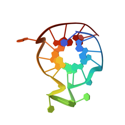
Solution structures of all parallel-stranded monomeric and dimeric G-quadruplex scaffolds of the human c-kit2 promoter.
Kuryavyi, V., Phan, A.T., Patel, D.J.(2010) Nucleic Acids Res 38: 6757-6773
- PubMed: 20566478 Search on PubMedSearch on PubMed Central
- DOI: https://doi.org/10.1093/nar/gkq558
- Primary Citation Related Structures:
2KYO, 2KYP - PubMed Abstract:
Previous studies have demonstrated that nuclease hypersensitivity regions of several proto-oncogenic DNA promoters, situated upstream of transcription start sites, contain guanine-rich tracts that form intramolecular G-quadruplexes stabilized by stacked G•G•G•G tetrads in monovalent cation solution. The human c-kit oncogenic promoter, an important target in the treatment of gastrointestinal tumors, contains two such stretches of guanine-rich tracts, designated c-kit1 and c-kit2. Our previous nuclear magnetic resonance (NMR)-based studies reported on the novel G-quadruplex scaffold of the c-kit1 promoter in K(+)-containing solution, where we showed for the first time that even an isolated guanine was involved in G-tetrad formation. These NMR-based studies are now extended to the c-kit2 promoter, which adopts two distinct all-parallel-stranded conformations in slow exchange, one of which forms a monomeric G-quadruplex (form-I) in 20 mM K(+)-containing solution and the other a novel dimeric G-quadruplex (form-II) in 100 mM K(+)-containing solution. The c-kit2 promoter dimeric form-II G-quadruplex adopts an unprecedented all-parallel-stranded topology where individual c-kit2 promoter strands span a pair of three-G-tetrad-layer-containing all-parallel-stranded G-quadruplexes aligned in a 3' to 5'-end orientation, with stacking continuity between G-quadruplexes mediated by a sandwiched A•A non-canonical pair. We propose that strand exchange during recombination events within guanine-rich segments, could potentially be mediated by a synapsis intermediate involving an intergenic parallel-stranded dimeric G-quadruplex.
- Memorial Sloan-Kettering Cancer Center, New York, NY 10065, USA.
Organizational Affiliation: