2KSV | pdb_00002ksv

The NMR structure of protein-glutaminase from Chryseobacterium proteolyticum


Experimental Data Snapshot

  • Method: SOLUTION NMR
  • Conformers Calculated: 100 
  • Conformers Submitted: 20 
  • Selection Criteria: target function 

wwPDB Validation 3D Report Full Report

Validation slider image for 2KSV

This is version 1.3 of the entry. See complete history

Literature

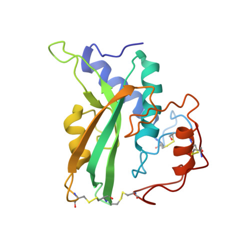
The NMR structure of protein-glutaminase from Chryseobacterium proteolyticum.

Kumeta, H.Miwa, N.Ogura, K.Kai, Y.Mizukoshi, T.Shimba, N.Suzuki, E.Inagaki, F.

(2010) J Biomol NMR 46: 251-255

Macromolecule Content 

  • Total Structure Weight: 19.88 kDa 
  • Atom Count: 1,388 
  • Modeled Residue Count: 185 
  • Deposited Residue Count: 185 
  • Unique protein chains: 1

Macromolecules

Find similar proteins by:|  3D Structure
Entity ID: 1
MoleculeChains  Sequence LengthOrganismDetailsImage
Protein-glutaminase185Chryseobacterium proteolyticumMutation(s): 0 
Gene Names: prgA
EC: 3.5.1.44
UniProt
Find proteins for Q9AQQ8 (Chryseobacterium proteolyticum)
Explore Q9AQQ8 
Go to UniProtKB:  Q9AQQ8
Entity Groups
Sequence Clusters30% Identity50% Identity70% Identity90% Identity95% Identity100% Identity
UniProt GroupQ9AQQ8
Sequence Annotations
Expand
Reference Sequence

Experimental Data & Validation

Experimental Data

  • Method: SOLUTION NMR
  • Conformers Calculated: 100 
  • Conformers Submitted: 20 
  • Selection Criteria: target function 

Structure Validation

View Full Validation Report


Entry History 

Deposition Data

Revision History  (Full details and data files)

  • Version 1.0: 2010-02-16
    Type: Initial release
  • Version 1.1: 2011-07-13
    Changes: Version format compliance
  • Version 1.2: 2022-03-16
    Changes: Data collection, Database references, Derived calculations
  • Version 1.3: 2024-10-30
    Changes: Data collection, Structure summary