2KSP | pdb_00002ksp

Mechanism for the selective interaction of C-terminal EH-domain proteins with specific NPF-containing partners


Experimental Data Snapshot

  • Method: SOLUTION NMR
  • Conformers Calculated: 30 
  • Conformers Submitted: 10 
  • Selection Criteria: structures with the lowest energy 

wwPDB Validation 3D Report Full Report

Validation slider image for 2KSP

This is version 1.3 of the entry. See complete history

Literature

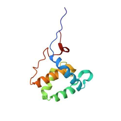
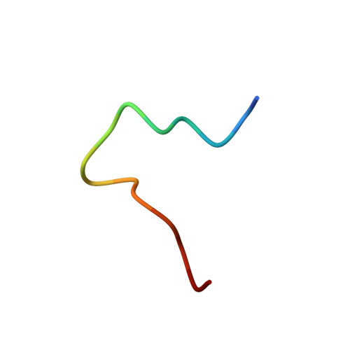
Mechanism for the selective interaction of C-terminal Eps15 homology domain proteins with specific Asn-Pro-Phe-containing partners.

Kieken, F.Sharma, M.Jovic, M.Giridharan, S.S.Naslavsky, N.Caplan, S.Sorgen, P.L.

(2010) J Biological Chem 285: 8687-8694

  • DOI: https://doi.org/10.1074/jbc.M109.045666
  • Primary Citation Related Structures: 
    2KSP

  • PubMed Abstract: 

    Epidermal growth factor receptor tyrosine kinase substrate 15 (Eps15) homology (EH)-domain proteins can be divided into two classes: those with an N-terminal EH-domain(s), and the C-terminal Eps15 homology domain-containing proteins (EHDs). Whereas many N-terminal EH-domain proteins regulate internalization events, the best characterized C-terminal EHD, EHD1, regulates endocytic recycling. Because EH-domains interact with the tripeptide Asn-Pro-Phe (NPF), it is of critical importance to elucidate the molecular mechanisms that allow EHD1 and its paralogs to interact selectively with a subset of the hundreds of NPF-containing proteins expressed in mammalian cells. Here, we capitalize on our findings that C-terminal EH-domains possess highly positively charged interaction surfaces and that many NPF-containing proteins that interact with C-terminal (but not N-terminal) EH-domains are followed by acidic residues. Using the recently identified EHD1 interaction partner molecule interacting with CasL (MICAL)-Like 1 (MICAL-L1) as a model, we have demonstrated that only the first of its two NPF motifs is required for EHD1 binding. Because only this first NPF is followed by acidic residues, we have utilized glutathione S-transferase pulldowns, two-hybrid analysis, and NMR to demonstrate that the flanking acidic residues "fine tune" the binding affinity to EHD1. Indeed, our NMR solution structure of the EHD1 EH-domain in complex with the MICAL-L1 NPFEEEEED peptide indicates that the first two flanking Glu residues lie in a position favorable to form salt bridges with Lys residues within the EH-domain. Our data provide a novel explanation for the selective interaction of C-terminal EH-domains with specific NPF-containing proteins and allow for the prediction of new interaction partners with C-terminal EHDs.


  • Organizational Affiliation
    • Department of Biochemistry and Molecular Biology and Eppley Cancer Center, University of Nebraska Medical Center, Omaha, Nebraska 68198-5870, USA.

Macromolecule Content 

  • Total Structure Weight: 13.55 kDa 
  • Atom Count: 954 
  • Modeled Residue Count: 120 
  • Deposited Residue Count: 120 
  • Unique protein chains: 2

Macromolecules

Find similar proteins by:|  3D Structure
Entity ID: 1
MoleculeChains  Sequence LengthOrganismDetailsImage
EH domain-containing protein 1105Homo sapiensMutation(s): 0 
Gene Names: EHD1PASTPAST1CDABP0131
UniProt & NIH Common Fund Data Resources
Find proteins for Q9H4M9 (Homo sapiens)
Explore Q9H4M9 
Go to UniProtKB:  Q9H4M9
PHAROS:  Q9H4M9
GTEx:  ENSG00000110047 
Entity Groups
Sequence Clusters30% Identity50% Identity70% Identity90% Identity95% Identity100% Identity
UniProt GroupQ9H4M9
Sequence Annotations
Expand
Reference Sequence
Find similar proteins by:  Sequence   |   3D Structure  
Entity ID: 2
MoleculeChains  Sequence LengthOrganismDetailsImage
MICAL L1 like peptide15Homo sapiensMutation(s): 0 
Gene Names: MICALL1
UniProt & NIH Common Fund Data Resources
Find proteins for Q8N3F8 (Homo sapiens)
Explore Q8N3F8 
Go to UniProtKB:  Q8N3F8
PHAROS:  Q8N3F8
GTEx:  ENSG00000100139 
Entity Groups
Sequence Clusters30% Identity50% Identity70% Identity90% Identity95% Identity100% Identity
UniProt GroupQ8N3F8
Sequence Annotations
Expand
Reference Sequence

Small Molecules

Ligands 1 Unique
IDChains Name / Formula / InChI Key2D Diagram3D Interactions
CA

Query on CA


Download:Ideal Coordinates CCD File
C [auth A]CALCIUM ION
Ca
BHPQYMZQTOCNFJ-UHFFFAOYSA-N

Experimental Data & Validation

Experimental Data

  • Method: SOLUTION NMR
  • Conformers Calculated: 30 
  • Conformers Submitted: 10 
  • Selection Criteria: structures with the lowest energy 

Structure Validation

View Full Validation Report


Entry History 

Deposition Data

Revision History  (Full details and data files)

  • Version 1.0: 2010-01-26
    Type: Initial release
  • Version 1.1: 2011-07-13
    Changes: Version format compliance
  • Version 1.2: 2020-02-26
    Changes: Database references, Derived calculations, Other
  • Version 1.3: 2024-05-01
    Changes: Data collection, Database references, Derived calculations