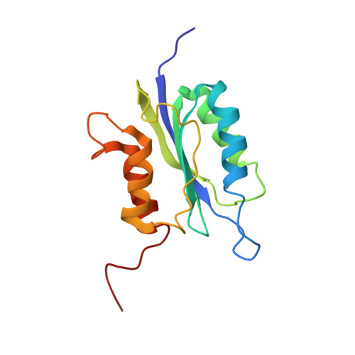
Structure and insight into blue light-induced changes in the BlrP1 BLUF domain
Wu, Q., Gardner, K.(2009) Biochemistry 48: 2620-2629
- PubMed: 19191473
- DOI: https://doi.org/10.1021/bi802237r
- Primary Citation of Related Structures:
2KB2 - PubMed Abstract:
BLUF domains (sensors of blue light using flavin adenine dinucleotide) are a group of flavin-containing blue light photosensory domains from a variety of bacterial and algal proteins. While spectroscopic studies have indicated that these domains reorganize their interactions with an internally bound chromophore upon illumination, it remains unclear how these are converted into structural and functional changes. To address this, we have solved the solution structure of the BLUF domain from Klebsiella pneumoniae BlrP1, a light-activated c-di-guanosine 5'-monophosphate phosphodiesterase which consists of a sensory BLUF and a catalytic EAL (Glu-Ala-Leu) domain [Schmidt et. al. (2008) J. Bacteriol. 187, 4774-4781]. Our dark state structure of the sensory domain shows that it adopts a standard BLUF domain fold followed by two C-terminal alpha helices which adopt a novel orientation with respect to the rest of the domain. Comparison of NMR spectra acquired under dark and light conditions suggests that residues throughout the BlrP1 BLUF domain undergo significant light-induced chemical shift changes, including sites clustered on the beta(4)beta(5) loop, beta(5) strand, and alpha(3)alpha(4) loop. Given that these changes were observed at several sites on the helical cap, over 15 A from chromophore, our data suggest a long-range signal transduction process in BLUF domains.
Organizational Affiliation:
Department of Biochemistry, University of Texas Southwestern Medical Center, Dallas, Texas 75390-8816, USA.


