2K6I | pdb_00002k6i

The domain features of the peripheral stalk subunit H of the methanogenic A1AO ATP synthase and the NMR solution structure of H1-47


Experimental Data Snapshot

  • Method: SOLUTION NMR
  • Conformers Calculated: 100 
  • Conformers Submitted: 10 
  • Selection Criteria: structures with the least restraint violations 

wwPDB Validation 3D Report Full Report

Validation slider image for 2K6I

This is version 1.4 of the entry. See complete history

Literature

Domain features of the peripheral stalk subunit H of the methanogenic A1AO ATP synthase and the NMR solution structure of H(1-47).

Biukovic, G.Gayen, S.Pervushin, K.Gruber, G.

(2009) Biophys J 97: 286-294

  • DOI: https://doi.org/10.1016/j.bpj.2009.04.026
  • Primary Citation Related Structures: 
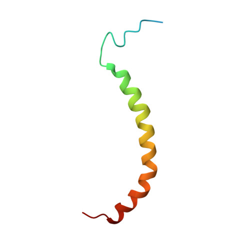
    2K6I

  • PubMed Abstract: 

    A series of truncated forms of subunit H were generated to establish the domain features of that protein. Circular dichroism analysis demonstrated that H is divided at least into a C-terminal coiled-coil domain within residues 54-104, and an N-terminal domain formed by adjacent alpha-helices. With a cysteine at the C-terminus of each of the truncated proteins (H(1-47), H(1-54), H(1-59), H(1-61), H(1-67), H(1-69), H(1-71), H(1-78), H(1-80), H(1-91), and H(47-105)), the residues involved in formation of the coiled-coil interface were determined. Proteins H(1-54), H(1-61), H(1-69), and H(1-80) showed strong cross-link formation, which was weaker in H(1-47), H(1-59), H(1-71), and H(1-91). A shift in disulfide formation between cysteines at positions 71 and 80 reflected an interruption in the periodicity of hydrophobic residues in the region 71AEKILEETEKE81. To understand how the N-terminal domain of H is formed, we determined for the first time, to our knowledge, the solution NMR structure of H(1-47), which revealed an alpha-helix between residues 15-42 and a flexible N-terminal stretch. The alpha-helix includes a kink that would bring the two helices of the C-terminus into the coiled-coil arrangement. H(1-47) revealed a strip of alanines involved in dimerization, which were tested by exchange to single cysteines in subunit H mutants.


  • Organizational Affiliation
    • School of Biological Sciences, Nanyang Technological University, Singapore.

Macromolecule Content 

  • Total Structure Weight: 6.39 kDa 
  • Atom Count: 360 
  • Modeled Residue Count: 47 
  • Deposited Residue Count: 56 
  • Unique protein chains: 1

Macromolecules

Find similar proteins by:|  3D Structure
Entity ID: 1
MoleculeChains  Sequence LengthOrganismDetailsImage
Uncharacterized protein MJ022356Methanocaldococcus jannaschiiMutation(s): 0 
Gene Names: MJ0223
UniProt
Find proteins for Q57676 (Methanocaldococcus jannaschii (strain ATCC 43067 / DSM 2661 / JAL-1 / JCM 10045 / NBRC 100440))
Explore Q57676 
Go to UniProtKB:  Q57676
Entity Groups
Sequence Clusters30% Identity50% Identity70% Identity90% Identity95% Identity100% Identity
UniProt GroupQ57676
Sequence Annotations
Expand
Reference Sequence

Experimental Data & Validation

Experimental Data

  • Method: SOLUTION NMR
  • Conformers Calculated: 100 
  • Conformers Submitted: 10 
  • Selection Criteria: structures with the least restraint violations 

Structure Validation

View Full Validation Report


Entry History 

Deposition Data

Revision History  (Full details and data files)

  • Version 1.0: 2009-07-21
    Type: Initial release
  • Version 1.1: 2011-07-13
    Changes: Version format compliance
  • Version 1.2: 2020-02-19
    Changes: Data collection, Database references, Derived calculations, Other
  • Version 1.3: 2023-06-14
    Changes: Database references, Other
  • Version 1.4: 2024-05-08
    Changes: Data collection, Database references