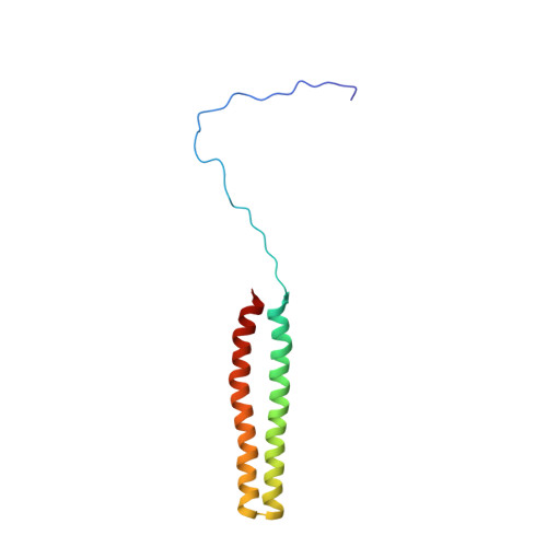
NMR Structure of the N-terminal Coiled Coil Domain of the Andes Hantavirus Nucleocapsid Protein.
Wang, Y., Boudreaux, D.M., Estrada, D.F., Egan, C.W., St Jeor, S.C., De Guzman, R.N.(2008) J Biological Chem 283: 28297-28304
- PubMed: 18687679 Search on PubMedSearch on PubMed Central
- DOI: https://doi.org/10.1074/jbc.M804869200
- Primary Citation Related Structures:
2K48 - PubMed Abstract:
The hantaviruses are emerging infectious viruses that in humans can cause a cardiopulmonary syndrome or a hemorrhagic fever with renal syndrome. The nucleocapsid (N) is the most abundant viral protein, and during viral assembly, the N protein forms trimers and packages the viral RNA genome. Here, we report the NMR structure of the N-terminal domain (residues 1-74, called N1-74) of the Andes hantavirus N protein. N1-74 forms two long helices (alpha1 and alpha2) that intertwine into a coiled coil domain. The conserved hydrophobic residues at the helix alpha1-alpha2 interface stabilize the coiled coil; however, there are many conserved surface residues whose function is not known. Site-directed mutagenesis, CD spectroscopy, and immunocytochemistry reveal that a point mutation in the conserved basic surface formed by Arg22 or Lys26 lead to antibody recognition based on the subcellular localization of the N protein. Thus, Arg22 and Lys26 are likely involved in a conformational change or molecular recognition when the N protein is trafficked from the cytoplasm to the Golgi, the site of viral assembly and maturation.
- Department of Molecular Biosciences, University of Kansas, Lawrence, Kansas 66045, USA.
Organizational Affiliation: