2K0U | pdb_00002k0u

High Resolution Solution NMR Structures of Oxaliplatin-DNA Adduct


Experimental Data Snapshot

  • Method: SOLUTION NMR
  • Conformers Calculated: 20 
  • Conformers Submitted: 
  • Selection Criteria: structures with the lowest energy 

wwPDB Validation 3D Report Full Report

Validation slider image for 2K0U

This is version 1.3 of the entry. See complete history

Literature

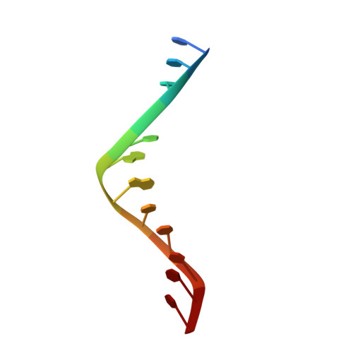
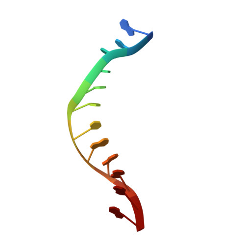
Flanking Bases Influence the Nature of DNA Distortion by Platinum 1,2-Intrastrand (GG) Cross-Links.

Bhattacharyya, D.Ramachandran, S.Sharma, S.Pathmasiri, W.King, C.L.Baskerville-Abraham, I.Boysen, G.Swenberg, J.A.Campbell, S.L.Dokholyan, N.V.Chaney, S.G.

(2011) PLoS One 6: e23582-e23582

  • DOI: https://doi.org/10.1371/journal.pone.0023582
  • Primary Citation Related Structures: 
    2K0T, 2K0U, 2K0V

  • PubMed Abstract: 

    The differences in efficacy and molecular mechanisms of platinum anti-cancer drugs cisplatin (CP) and oxaliplatin (OX) are thought to be partially due to the differences in the DNA conformations of the CP and OX adducts that form on adjacent guanines on DNA, which in turn influence the binding of damage-recognition proteins that control downstream effects of the adducts. Here we report a comprehensive comparison of the structural distortion of DNA caused by CP and OX adducts in the TGGT sequence context using nuclear magnetic resonance (NMR) spectroscopy and molecular dynamics (MD) simulations. When compared to our previous studies in other sequence contexts, these structural studies help us understand the effect of the sequence context on the conformation of Pt-GG DNA adducts. We find that both the sequence context and the type of Pt-GG DNA adduct (CP vs. OX) play an important role in the conformation and the conformational dynamics of Pt-DNA adducts, possibly explaining their influence on the ability of many damage-recognition proteins to bind to Pt-DNA adducts.


  • Organizational Affiliation
    • Department of Biochemistry and Biophysics, School of Medicine, University of North Carolina, Chapel Hill, North Carolina, United States of America.

Macromolecule Content 

  • Total Structure Weight: 7.64 kDa 
  • Atom Count: 495 
  • Modeled Residue Count: 24 
  • Deposited Residue Count: 24 
  • Unique nucleic acid chains: 2

Macromolecules

Find similar nucleic acids by:  Sequence
Entity ID: 1
MoleculeChains LengthOrganismImage
DNA (5'-D(*DCP*DCP*DTP*DCP*DTP*DGP*DGP*DTP*DCP*DTP*DCP*DC)-3')12N/A
Sequence Annotations
Expand
Reference Sequence
Find similar nucleic acids by:  Sequence
Entity ID: 2
MoleculeChains LengthOrganismImage
DNA (5'-D(*DGP*DGP*DAP*DGP*DAP*DCP*DCP*DAP*DGP*DAP*DGP*DG)-3')12N/A
Sequence Annotations
Expand
Reference Sequence

Small Molecules

Ligands 1 Unique
IDChains Name / Formula / InChI Key2D Diagram3D Interactions
1PT

Query on 1PT


Download:Ideal Coordinates CCD File
C [auth A]CYCLOHEXANE-1(R),2(R)-DIAMINE-PLATINUM(II)
C6 H14 N2 Pt
SGLJYTWMWIAGEU-KGZKBUQUSA-N

Experimental Data & Validation

Experimental Data

  • Method: SOLUTION NMR
  • Conformers Calculated: 20 
  • Conformers Submitted: 
  • Selection Criteria: structures with the lowest energy 

Structure Validation

View Full Validation Report


Entry History 

Deposition Data

Revision History  (Full details and data files)

  • Version 1.0: 2009-02-03
    Type: Initial release
  • Version 1.1: 2011-07-13
    Changes: Version format compliance
  • Version 1.2: 2011-08-31
    Changes: Database references
  • Version 1.3: 2024-05-01
    Changes: Data collection, Database references, Derived calculations