Solution NMR Structure of Selenium-binding Protein from Methanococcus vannielii.
Suzuki, M., Lee, D.Y., Inyamah, N., Stadtman, T.C., Tjandra, N.(2008) J Biological Chem 283: 25936-25943
- PubMed: 18650445 Search on PubMedSearch on PubMed Central
- DOI: https://doi.org/10.1074/jbc.M803773200
- Primary Citation Related Structures:
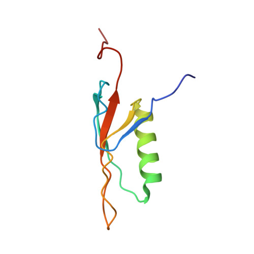
2JZ7 - PubMed Abstract:
Selenium is an important nutrient. The lack of selenium will suppress expression of various enzymes that will lead to cell abnormality and diseases. However, high concentrations of free selenium are toxic to the cell because it adversely affects numerous cell metabolic pathways. In Methanococcus vannielii, selenium transport in the cell is established by the selenium-binding protein, SeBP. SeBP sequesters selenium during transport, thus regulating the level of free selenium in the cell, and delivers it specifically to the selenophosphate synthase enzyme. In solution, SeBP is an oligomer of 8.8-kDa subunits. It is a symmetric pentamer. The solution structure of SeBP was determined by NMR spectroscopy. Each subunit of SeBP is composed of an alpha-helix on top of a 4-stranded twisted beta-sheet. The stability of the five subunits stems mainly from hydrophobic interactions and supplemented by hydrogen bond interactions. The loop containing Cys(59) has been shown to be important for selenium binding, is flexible, and adopts multiple conformations. However, the cysteine accessibility is restricted in the structure, reducing the possibility of the binding of free selenium readily. Therefore, a different selenium precursor or other factors might be needed to facilitate opening of this loop to expose Cys(59) for selenium binding.
- Laboratory of Molecular Biophysics, NHLBI, National Institutes of Health, Bethesda, Maryland 20892, USA.
Organizational Affiliation: