Interaction between the N-terminal SH3 domain of Nckalpha and CD3epsilon-derived peptides: Non-canonical and canonical recognition motifs
Santiveri, C.M., Borroto, A., Simon, L., Rico, M., Alarcon, B., Jimenez, M.A.(2009) Biochim Biophys Acta Proteins Proteom 1794: 110-117
- PubMed: 18955169 Search on PubMed
- DOI: https://doi.org/10.1016/j.bbapap.2008.09.016
- Primary Citation Related Structures:
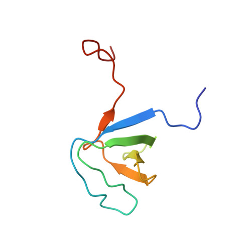
2JW4 - PubMed Abstract:
The first SH3 domain (SH3.1) of Nckalpha specifically recognizes the proline-rich region of CD3varepsilon, a subunit of the T cell receptor complex. We have solved the NMR structure of Nckalpha SH3.1 that shows the characteristic SH3 fold consisting of two antiparallel beta-sheets tightly packed against each other. According to chemical shift mapping analysis, a peptide encompassing residues 150-166 of CD3varepsilon binds at the canonical SH3 binding site. An exhaustive comparison with the structures of other SH3 domains able and unable to bind CD3varepsilon reveals that Nckalpha SH3.1 recognises a non-canonical PxxPxxDY motif that orientates at the binding site as a class II ligand. A positively charged residue (K/R) at position -2 relative to the WW sequence at the beginning of strand beta3 is crucial for PxxDY recognition. A 14-mer optimised Nckalpha SH3.1 ligand was found using a multi-substitution approach. Based on NMR data, this improved ligand binds Nckalpha SH3.1 through a PxxPxRDY motif that combines specific stabilising interactions corresponding to both canonical class II, PxxPx(K/R), and non-canonical PxxPxxDY motifs. This explains its higher capacity for Nckalpha SH3.1 binding relative to the wild type sequence.
- Instituto de Química Física Rocasolano, CSIC, Serrano-119, 28006-Madrid, Spain.
Organizational Affiliation: