2JJB | pdb_00002jjb

Family 37 trehalase from Escherichia coli in complex with casuarine-6- O-alpha-glucopyranose


Experimental Data Snapshot

  • Method: X-RAY DIFFRACTION
  • Resolution: 1.90 Å
  • R-Value Free: 
    0.224 (Depositor), 0.224 (DCC) 
  • R-Value Work: 
    0.177 (Depositor), 0.177 (DCC) 
  • R-Value Observed: 
    0.179 (Depositor) 

Starting Model: experimental
View more details

wwPDB Validation 3D Report Full Report

Validation slider image for 2JJB

Ligand Structure Quality Assessment 


This is version 1.5 of the entry. See complete history

Literature

Total Syntheses of Casuarine and its 6-O-Alpha-Glucoside: Complementary Inhibition Towards Glycoside Hydrolases of the Gh31 and Gh37 Families.

Cardona, F.Parmeggiani, C.Faggi, E.Bonaccini, C.Gratteri, P.Sim, L.Gloster, T.M.Roberts, S.Davies, G.J.Rose, D.R.Goti, A.

(2009) Chemistry 15: 1627

  • DOI: https://doi.org/10.1002/chem.200801578
  • Primary Citation Related Structures: 
    2JJB, 3CTT

  • PubMed Abstract: 

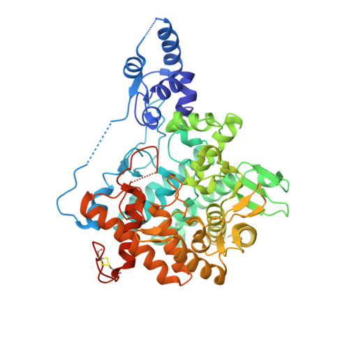
    Total synthesis of naturally occurring casuarine (1) and the first total synthesis of casuarine 6-O-alpha-glucoside (2) were achieved through complete stereoselective nitrone cycloaddition, Tamao-Fleming oxidation and selective alpha-glucosylation as key steps. Biological assays of the two compounds proved their strong and selective inhibitory properties towards glucoamylase NtMGAM and trehalase Tre37A, respectively, which place them among the most powerful inhibitors of these enzymes. The structural determination of the complexes of NtMGAM with 1 and of Tre37A with 2 revealed interesting similarities in the catalytic sites of these two enzymes which belong to different families and clans.


  • Organizational Affiliation
    • Department of Organic Chemistry U. Schiff, Laboratory of Design, Synthesis and Study of Biologically Active Heterocycles, University of Florence, Via della Lastruccia, 13, 50019 Sesto Fiorentino (FI), Italy.

Macromolecule Content 

  • Total Structure Weight: 247.48 kDa 
  • Atom Count: 18,411 
  • Modeled Residue Count: 1,997 
  • Deposited Residue Count: 2,140 
  • Unique protein chains: 1

Macromolecules

Find similar proteins by:|  3D Structure
Entity ID: 1
MoleculeChains  Sequence LengthOrganismDetailsImage
PERIPLASMIC TREHALASE
A, B, C, D
535Escherichia coli K-12Mutation(s): 0 
EC: 3.2.1.28
UniProt
Find proteins for P13482 (Escherichia coli (strain K12))
Explore P13482 
Go to UniProtKB:  P13482
Entity Groups
Sequence Clusters30% Identity50% Identity70% Identity90% Identity95% Identity100% Identity
UniProt GroupP13482
Sequence Annotations
Expand
Reference Sequence

Small Molecules

Ligands 4 Unique
IDChains Name / Formula / InChI Key2D Diagram3D Interactions
3CU

Query on 3CU


Download:Ideal Coordinates CCD File
DA [auth C],
E [auth A],
RA [auth D],
T [auth B]
CASUARINE
C8 H15 N O5
AXTGOJVKRHFYBT-XAZAIFFQSA-N
GLC

Query on GLC


Download:Ideal Coordinates CCD File
EA [auth C],
F [auth A],
SA [auth D],
U [auth B]
alpha-D-glucopyranose
C6 H12 O6
WQZGKKKJIJFFOK-DVKNGEFBSA-N
SO4

Query on SO4


Download:Ideal Coordinates CCD File
AA [auth B]
AB [auth D]
BA [auth B]
FA [auth C]
G [auth A]
AA [auth B],
AB [auth D],
BA [auth B],
FA [auth C],
G [auth A],
GA [auth C],
H [auth A],
HA [auth C],
I [auth A],
IA [auth C],
J [auth A],
JA [auth C],
K [auth A],
KA [auth C],
L [auth A],
LA [auth C],
M [auth A],
MA [auth C],
N [auth A],
NA [auth C],
O [auth A],
OA [auth C],
P [auth A],
PA [auth C],
Q [auth A],
R [auth A],
TA [auth D],
UA [auth D],
V [auth B],
VA [auth D],
W [auth B],
WA [auth D],
X [auth B],
XA [auth D],
Y [auth B],
YA [auth D],
Z [auth B],
ZA [auth D]
SULFATE ION
O4 S
QAOWNCQODCNURD-UHFFFAOYSA-L
EDO

Query on EDO


Download:Ideal Coordinates CCD File
CA [auth B],
QA [auth C],
S [auth A]
1,2-ETHANEDIOL
C2 H6 O2
LYCAIKOWRPUZTN-UHFFFAOYSA-N

Experimental Data & Validation

Experimental Data

  • Method: X-RAY DIFFRACTION
  • Resolution: 1.90 Å
  • R-Value Free:  0.224 (Depositor), 0.224 (DCC) 
  • R-Value Work:  0.177 (Depositor), 0.177 (DCC) 
  • R-Value Observed: 0.179 (Depositor) 
Space Group: P 21 21 21
Unit Cell:
Length ( Å )Angle ( ˚ )
a = 92.481α = 90
b = 117.863β = 90
c = 203.451γ = 90
Software Package:
Software NamePurpose
REFMACrefinement
DENZOdata reduction
SCALEPACKdata scaling
PHASERphasing

Structure Validation

View Full Validation Report


Ligand Structure Quality Assessment 


Entry History 

Deposition Data

Revision History  (Full details and data files)

  • Version 1.0: 2009-01-13
    Type: Initial release
  • Version 1.1: 2011-05-07
    Changes: Version format compliance
  • Version 1.2: 2011-07-13
    Changes: Version format compliance
  • Version 1.3: 2020-07-29
    Type: Remediation
    Reason: Carbohydrate remediation
    Changes: Data collection, Derived calculations, Other, Structure summary
  • Version 1.4: 2023-12-13
    Changes: Data collection, Database references, Refinement description, Structure summary
  • Version 1.5: 2024-11-20
    Changes: Structure summary