2JIG | pdb_00002jig

Crystal structure of Chlamydomonas reinhardtii prolyl-4 hydroxylase type I complexed with zinc and pyridine-2,4-dicarboxylate


Experimental Data Snapshot

  • Method: X-RAY DIFFRACTION
  • Resolution: 1.85 Å
  • R-Value Free: 
    0.238 (Depositor), 0.236 (DCC) 
  • R-Value Work: 
    0.183 (Depositor), 0.184 (DCC) 
  • R-Value Observed: 
    0.186 (Depositor) 

Starting Model: experimental
View more details

wwPDB Validation 3D Report Full Report

Validation slider image for 2JIG

Ligand Structure Quality Assessment 


This is version 1.4 of the entry. See complete history

Literature

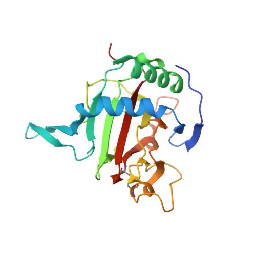
The Active Site of an Algal Prolyl 4-Hydroxylase Has a Large Structural Plasticity.

Koski, M.K.Hieta, R.Bollner, C.Kivirikko, K.I.Myllyharju, J.Wierenga, R.K.

(2007) J Biological Chem 282: 37112

  • DOI: https://doi.org/10.1074/jbc.M706554200
  • Primary Citation Related Structures: 
    2JIG, 2JIJ, 2V4A

  • PubMed Abstract: 

    Prolyl 4-hydroxylases (P4Hs) are 2-oxoglutarate dioxygenases that catalyze the hydroxylation of peptidyl prolines. They play an important role in collagen synthesis, oxygen homeostasis, and plant cell wall formation. We describe four structures of a P4H from the green alga Chlamydomonas reinhardtii, two of the apoenzyme at 1.93 and 2.90 A resolution, one complexed with the competitive inhibitor Zn2+, and one with Zn2+ and pyridine 2,4-dicarboxylate (which is an analogue of 2-oxoglutarate) at 1.85 A resolution. The structures reveal the double-stranded beta-helix core fold (jellyroll motif), typical for 2-oxoglutarate dioxygenases. The catalytic site is at the center of an extended shallow groove lined by two flexible loops. Mutagenesis studies together with the crystallographic data indicate that this groove participates in the binding of the proline-rich peptide-substrates. It is discussed that the algal P4H and the catalytic domain of collagen P4Hs have notable structural similarities, suggesting that these enzymes form a separate structural subgroup of P4Hs different from the hypoxia-inducible factor P4Hs. Key structural differences between these two subgroups are described. These studies provide first insight into the structure-function relationships of the collagen P4Hs, which unlike the hypoxia-inducible factor P4Hs use proline-rich peptides as their substrates.


  • Organizational Affiliation
    • Biocenter Oulu and Department of Biochemistry and Collagen Research Unit, University of Oulu, FIN-90014 Oulu, Finland.

Macromolecule Content 

  • Total Structure Weight: 50.54 kDa 
  • Atom Count: 3,687 
  • Modeled Residue Count: 411 
  • Deposited Residue Count: 448 
  • Unique protein chains: 1

Macromolecules

Find similar proteins by:|  3D Structure
Entity ID: 1
MoleculeChains  Sequence LengthOrganismDetailsImage
PROLYL-4 HYDROXYLASE
A, B
224Chlamydomonas reinhardtiiMutation(s): 0 

Small Molecules

Ligands 4 Unique
IDChains Name / Formula / InChI Key2D Diagram3D Interactions
PD2

Query on PD2


Download:Ideal Coordinates CCD File
E [auth A]PYRIDINE-2,4-DICARBOXYLIC ACID
C7 H5 N O4
MJIVRKPEXXHNJT-UHFFFAOYSA-N
SO4

Query on SO4


Download:Ideal Coordinates CCD File
G [auth A]SULFATE ION
O4 S
QAOWNCQODCNURD-UHFFFAOYSA-L
GOL

Query on GOL


Download:Ideal Coordinates CCD File
F [auth A]GLYCEROL
C3 H8 O3
PEDCQBHIVMGVHV-UHFFFAOYSA-N
ZN

Query on ZN


Download:Ideal Coordinates CCD File
C [auth A],
D [auth A],
H [auth B],
I [auth B]
ZINC ION
Zn
PTFCDOFLOPIGGS-UHFFFAOYSA-N

Experimental Data & Validation

Experimental Data

  • Method: X-RAY DIFFRACTION
  • Resolution: 1.85 Å
  • R-Value Free:  0.238 (Depositor), 0.236 (DCC) 
  • R-Value Work:  0.183 (Depositor), 0.184 (DCC) 
  • R-Value Observed: 0.186 (Depositor) 
Space Group: P 21 21 21
Unit Cell:
Length ( Å )Angle ( ˚ )
a = 57.92α = 90
b = 60.25β = 90
c = 116.85γ = 90
Software Package:
Software NamePurpose
REFMACrefinement
XDSdata reduction
XDSdata scaling
PHASERphasing

Structure Validation

View Full Validation Report


Ligand Structure Quality Assessment 


Entry History 

Deposition Data

Revision History  (Full details and data files)

  • Version 1.0: 2007-10-30
    Type: Initial release
  • Version 1.1: 2011-07-13
    Changes: Advisory, Refinement description, Version format compliance
  • Version 1.2: 2019-07-24
    Changes: Data collection
  • Version 1.3: 2023-12-13
    Changes: Data collection, Database references, Derived calculations, Other, Refinement description
  • Version 1.4: 2024-10-23
    Changes: Structure summary