2JHQ | pdb_00002jhq

Crystal structure of Uracil DNA-glycosylase from Vibrio cholerae


Experimental Data Snapshot

  • Method: X-RAY DIFFRACTION
  • Resolution: 1.50 Å
  • R-Value Free: 
    0.210 (Depositor), 0.209 (DCC) 
  • R-Value Work: 
    0.175 (Depositor), 0.175 (DCC) 
  • R-Value Observed: 
    0.177 (Depositor) 

Starting Model: experimental
View more details

wwPDB Validation 3D Report Full Report

Validation slider image for 2JHQ

This is version 1.2 of the entry. See complete history

Literature

Structure of Uracil-DNA N-Glycosylase (Ung) from Vibrio Cholerae. Mapping Temperature Adaptation Through Structural and Mutational Analysis.

Raeder, I.L.U.Moe, E.Willassen, N.P.Smalas, A.O.Leiros, I.

(2010) Acta Crystallogr Sect F Struct Biol Cryst Commun 66: 130

  • DOI: https://doi.org/10.1107/S1744309109052063
  • Primary Citation Related Structures: 
    2JHQ

  • PubMed Abstract: 

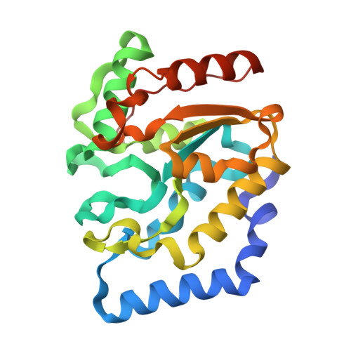
    The crystal structure of Vibrio cholerae uracil-DNA N-glycosylase (vcUNG) has been determined to 1.5 A resolution. Based on this structure, a homology model of Aliivibrio salmonicida uracil-DNA N-glycosylase (asUNG) was built. A previous study demonstrated that asUNG possesses typical cold-adapted features compared with vcUNG, such as a higher catalytic efficiency owing to increased substrate affinity. Specific amino-acid substitutions in asUNG were suggested to be responsible for the increased substrate affinity and the elevated catalytic efficiency by increasing the positive surface charge in the DNA-binding region. The temperature adaptation of these enzymes has been investigated using structural and mutational analyses, in which mutations of vcUNG demonstrated an increased substrate affinity that more resembled that of asUNG. Visualization of surface potentials revealed a more positive potential for asUNG compared with vcUNG; a modelled double mutant of vcUNG had a potential around the substrate-binding region that was more like that of asUNG, thus rationalizing the results obtained from the kinetic studies.


  • Organizational Affiliation
    • Department of Chemistry, Faculty of Science, University of Tromsø, N-9037 Tromsø, Norway.

Macromolecule Content 

  • Total Structure Weight: 25.51 kDa 
  • Atom Count: 2,122 
  • Modeled Residue Count: 223 
  • Deposited Residue Count: 226 
  • Unique protein chains: 1

Macromolecules

Find similar proteins by:|  3D Structure
Entity ID: 1
MoleculeChains  Sequence LengthOrganismDetailsImage
URACIL DNA-GLYCOSYLASE226Vibrio choleraeMutation(s): 0 
EC: 3.2.2.3 (PDB Primary Data), 3.2.2.27 (UniProt)
UniProt
Find proteins for Q9KPK8 (Vibrio cholerae serotype O1 (strain ATCC 39315 / El Tor Inaba N16961))
Explore Q9KPK8 
Go to UniProtKB:  Q9KPK8
Entity Groups
Sequence Clusters30% Identity50% Identity70% Identity90% Identity95% Identity100% Identity
UniProt GroupQ9KPK8
Sequence Annotations
Expand
Reference Sequence

Small Molecules

Ligands 1 Unique
IDChains Name / Formula / InChI Key2D Diagram3D Interactions
CL

Query on CL


Download:Ideal Coordinates CCD File
B [auth A]CHLORIDE ION
Cl
VEXZGXHMUGYJMC-UHFFFAOYSA-M

Experimental Data & Validation

Experimental Data

  • Method: X-RAY DIFFRACTION
  • Resolution: 1.50 Å
  • R-Value Free:  0.210 (Depositor), 0.209 (DCC) 
  • R-Value Work:  0.175 (Depositor), 0.175 (DCC) 
  • R-Value Observed: 0.177 (Depositor) 
Space Group: P 21 21 21
Unit Cell:
Length ( Å )Angle ( ˚ )
a = 60.14α = 90
b = 60.44β = 90
c = 61.33γ = 90
Software Package:
Software NamePurpose
REFMACrefinement
XDSdata reduction
XDSdata scaling
MOLREPphasing

Structure Validation

View Full Validation Report


Entry History 

Deposition Data

Revision History  (Full details and data files)

  • Version 1.0: 2008-06-03
    Type: Initial release
  • Version 1.1: 2012-04-04
    Changes: Database references, Derived calculations, Non-polymer description, Structure summary, Version format compliance
  • Version 1.2: 2023-12-13
    Changes: Data collection, Database references, Derived calculations, Other, Refinement description