2JH3 | pdb_00002jh3

The crystal structure of DR2241 from Deinococcus radiodurans at 1.9 A resolution reveals a multi-domain protein with structural similarity to chelatases but also with two additional novel domains


Experimental Data Snapshot

  • Method: X-RAY DIFFRACTION
  • Resolution: 1.90 Å
  • R-Value Free: 
    0.238 (Depositor), 0.245 (DCC) 
  • R-Value Work: 
    0.189 (Depositor), 0.197 (DCC) 
  • R-Value Observed: 
    0.190 (Depositor) 

wwPDB Validation 3D Report Full Report

Validation slider image for 2JH3

This is version 1.3 of the entry. See complete history

Literature

The Crystal Structure of Dr2241 from Deinococcus Radiodurans at 1.9 A Resolution Reveals a Multi-Domain Protein with Structural Similarity to Chelatases But Also with Two Additional Novel Domains

Leiros, H.-K.S.Mcsweeney, S.M.

(2007) J Struct Biol 159: 92

  • DOI: https://doi.org/10.1016/j.jsb.2007.02.009
  • Primary Citation Related Structures: 
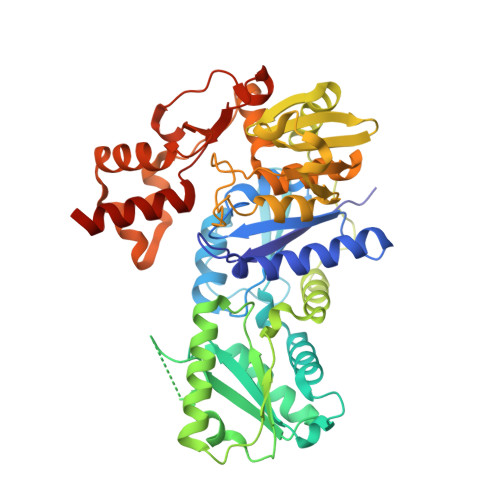
    2JH3

  • PubMed Abstract: 

    A unique family of proteins have been identified in the Deinococcus genus with an N-terminal cobalamin (vitamin B(12)) chelatase domain denoted CbiX and an additional unique C-terminal domain with unknown function. Here we report the first crystal structure from this new family of proteins with the structure of Deinococcus radiodurans protein DR2241. The structure reveals a multi-domain protein where domains A (residues 1-132) has the same fold as the small CbiX (CbiX(S)), domains A and B (residues 1-272) follow the chelatase super-family fold and the two additional unique domains C and D have no structural homologues. Domain D harbours the sequence motifs CxxC and CxxxC, in which DR2241 gives the first evidence that these motifs bind a [4Fe-4S] iron-sulphur cluster. In solution there are indications of multimeric forms, and in the crystallographic asymmetric unit a tetramer is found where domains C and D are involved in stabilising the tetrameric assembly.


  • Organizational Affiliation
    • Macromolecular Crystallography Group, European Synchrotron Radiation Facility (ESRF), BP 220, 6, Rue Jules Horowitz, F-38043 Grenoble Cedex 09, France.

Macromolecule Content 

  • Total Structure Weight: 205.68 kDa 
  • Atom Count: 14,998 
  • Modeled Residue Count: 1,833 
  • Deposited Residue Count: 1,896 
  • Unique protein chains: 1

Macromolecules

Find similar proteins by:|  3D Structure
Entity ID: 1
MoleculeChains  Sequence LengthOrganismDetailsImage
RIBOSOMAL PROTEIN S2-RELATED PROTEIN
A, B, C, D
474Deinococcus radioduransMutation(s): 0 
UniProt
Find proteins for Q9RS86 (Deinococcus radiodurans (strain ATCC 13939 / DSM 20539 / JCM 16871 / CCUG 27074 / LMG 4051 / NBRC 15346 / NCIMB 9279 / VKM B-1422 / R1))
Explore Q9RS86 
Go to UniProtKB:  Q9RS86
Entity Groups
Sequence Clusters30% Identity50% Identity70% Identity90% Identity95% Identity100% Identity
UniProt GroupQ9RS86
Sequence Annotations
Expand
Reference Sequence

Experimental Data & Validation

Experimental Data

  • Method: X-RAY DIFFRACTION
  • Resolution: 1.90 Å
  • R-Value Free:  0.238 (Depositor), 0.245 (DCC) 
  • R-Value Work:  0.189 (Depositor), 0.197 (DCC) 
  • R-Value Observed: 0.190 (Depositor) 
Space Group: P 21 21 21
Unit Cell:
Length ( Å )Angle ( ˚ )
a = 124.78α = 90
b = 128.69β = 90
c = 135.08γ = 90
Software Package:
Software NamePurpose
XDSdata reduction
XDSdata scaling
SHARPphasing
SOLVEphasing
REFMACrefinement

Structure Validation

View Full Validation Report


Entry History 

Deposition Data

Revision History  (Full details and data files)

  • Version 1.0: 2007-06-19
    Type: Initial release
  • Version 1.1: 2011-05-08
    Changes: Version format compliance
  • Version 1.2: 2011-07-13
    Changes: Version format compliance
  • Version 1.3: 2024-05-08
    Changes: Data collection, Database references, Derived calculations, Other