2JFS | pdb_00002jfs

Crystal structure of the PPM Ser-Thr phosphatase MsPP from Mycobacterium smegmatis in complex with cacodylate


Experimental Data Snapshot

  • Method: X-RAY DIFFRACTION
  • Resolution: 1.45 Å
  • R-Value Free: 
    0.191 (Depositor), 0.189 (DCC) 
  • R-Value Work: 
    0.178 (Depositor), 0.176 (DCC) 
  • R-Value Observed: 
    0.179 (Depositor) 

Starting Model: experimental
View more details

wwPDB Validation 3D Report Full Report

Validation slider image for 2JFS

This is version 1.2 of the entry. See complete history

Literature

Insights Into the Mechanism of Ppm Ser/Thr Phosphatases from the Atomic Resolution Structures of a Mycobacterial Enzyme

Bellinzoni, M.Wehenkel, A.Shepard, W.Alzari, P.M.

(2007) Structure 15: 863

  • DOI: https://doi.org/10.1016/j.str.2007.06.002
  • Primary Citation Related Structures: 
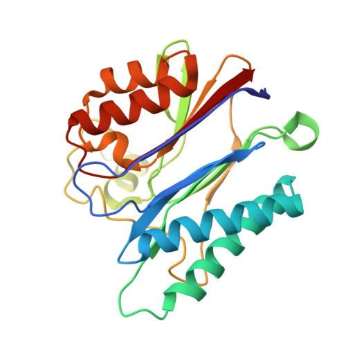
    2JFR, 2JFS, 2JFT

  • PubMed Abstract: 

    Serine/threonine-specific phosphatases (PPs) represent, after protein tyrosine phosphatases, the second major class of enzymes that catalyze the dephosphorylation of proteins. They are classed in two large families, known as PPP and PPM, on the basis of sequence similarities, metal ion dependence, and inhibitor sensitivity. Despite their wide species distribution and broad physiological roles, the catalytic mechanism of PPM phosphatases has been primarily inferred from studies of a single enzyme, human PP2Calpha. Here, we report the biochemical characterization and the atomic resolution structures of a soluble PPM phosphatase from the saprophyte Mycobacterium smegmatis in complex with different ligands. The structures provide putative snapshots along the catalytic cycle, which support an associative reaction mechanism that differs in some important aspects from the currently accepted model and reinforces the hypothesis of convergent evolution in PPs.


  • Organizational Affiliation
    • Unité de Biochimie Structurale, CNRS-URA 2185, Institut Pasteur, 75724 Paris Cedex 15, France.

Macromolecule Content 

  • Total Structure Weight: 24.86 kDa 
  • Atom Count: 2,056 
  • Modeled Residue Count: 232 
  • Deposited Residue Count: 234 
  • Unique protein chains: 1

Macromolecules

Find similar proteins by:|  3D Structure
Entity ID: 1
MoleculeChains  Sequence LengthOrganismDetailsImage
SER-THR PHOSPHATASE MSPP234Mycolicibacterium smegmatis MC2 155Mutation(s): 0 
UniProt
Find proteins for A0QTQ6 (Mycolicibacterium smegmatis (strain ATCC 700084 / mc(2)155))
Explore A0QTQ6 
Go to UniProtKB:  A0QTQ6
Entity Groups
Sequence Clusters30% Identity50% Identity70% Identity90% Identity95% Identity100% Identity
UniProt GroupA0QTQ6
Sequence Annotations
Expand
Reference Sequence

Small Molecules

Ligands 3 Unique
IDChains Name / Formula / InChI Key2D Diagram3D Interactions
CAC

Query on CAC


Download:Ideal Coordinates CCD File
E [auth A]CACODYLATE ION
C2 H6 As O2
OGGXGZAMXPVRFZ-UHFFFAOYSA-M
GOL

Query on GOL


Download:Ideal Coordinates CCD File
F [auth A],
G [auth A],
H [auth A]
GLYCEROL
C3 H8 O3
PEDCQBHIVMGVHV-UHFFFAOYSA-N
MN

Query on MN


Download:Ideal Coordinates CCD File
B [auth A],
C [auth A],
D [auth A]
MANGANESE (II) ION
Mn
WAEMQWOKJMHJLA-UHFFFAOYSA-N

Experimental Data & Validation

Experimental Data

  • Method: X-RAY DIFFRACTION
  • Resolution: 1.45 Å
  • R-Value Free:  0.191 (Depositor), 0.189 (DCC) 
  • R-Value Work:  0.178 (Depositor), 0.176 (DCC) 
  • R-Value Observed: 0.179 (Depositor) 
Space Group: P 21 21 21
Unit Cell:
Length ( Å )Angle ( ˚ )
a = 36.568α = 90
b = 65.786β = 90
c = 110.059γ = 90
Software Package:
Software NamePurpose
REFMACrefinement
MOSFLMdata reduction
SCALAdata scaling
AMoREphasing

Structure Validation

View Full Validation Report


Entry History 

Deposition Data

Revision History  (Full details and data files)

  • Version 1.0: 2007-07-24
    Type: Initial release
  • Version 1.1: 2011-07-13
    Changes: Advisory, Version format compliance
  • Version 1.2: 2023-12-13
    Changes: Data collection, Database references, Derived calculations, Other, Refinement description