2JCP | pdb_00002jcp

The hyaluronan binding domain of murine CD44


Experimental Data Snapshot

  • Method: X-RAY DIFFRACTION
  • Resolution: 1.30 Å
  • R-Value Free: 
    0.191 (Depositor), 0.192 (DCC) 
  • R-Value Work: 
    0.157 (Depositor), 0.160 (DCC) 
  • R-Value Observed: 
    0.158 (Depositor) 

Starting Model: experimental
View more details

wwPDB Validation 3D Report Full Report

Validation slider image for 2JCP

This is version 1.6 of the entry. See complete history

Literature

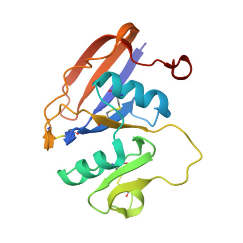
Structures of the Cd44-Hyaluronan Complex Provide Insight Into a Fundamental Carbohydrate-Protein Interaction.

Banerji, S.Wright, A.J.Noble, M.E.M.Mahoney, D.J.Campbell, I.D.Day, A.J.Jackson, D.G.

(2008) Nat Struct Mol Biol 14: 234

  • DOI: https://doi.org/10.1038/nsmb1201
  • Primary Citation Related Structures: 
    2JCP, 2JCQ, 2JCR

  • PubMed Abstract: 

    Regulation of transient interactions between cells and the ubiquitous matrix glycosaminoglycan hyaluronan is crucial to such fundamental processes as embryonic development and leukocyte homing. Cd44, the primary cell surface receptor for hyaluronan, binds ligand via a lectin-like fold termed the Link module, but only after appropriate functional activation. The molecular details of the Cd44-hyaluronan interaction and hence the structural basis for this activation are unknown. Here we present the first crystal structure of Cd44 complexed with hyaluronan. This reveals that the interaction with hyaluronan is dominated by shape and hydrogen-bonding complementarity and identifies two conformational forms of the receptor that differ in orientation of a crucial hyaluronan-binding residue (Arg45, equivalent to Arg41 in human CD44). Measurements by NMR indicate that the conformational transition can be induced by hyaluronan binding, providing further insight into possible mechanisms for regulation of Cd44.


  • Organizational Affiliation
    • Medical Research Council Human Immunology Unit, Weatherall Institute of Molecular Medicine, John Radcliffe Hospital, Headington, Oxford OX3 9DS, UK.

Macromolecule Content 

  • Total Structure Weight: 17.14 kDa 
  • Atom Count: 1,424 
  • Modeled Residue Count: 150 
  • Deposited Residue Count: 154 
  • Unique protein chains: 1

Macromolecules

Find similar proteins by:|  3D Structure
Entity ID: 1
MoleculeChains  Sequence LengthOrganismDetailsImage
CD44 ANTIGEN154Mus musculusMutation(s): 0 
UniProt
Find proteins for P15379 (Mus musculus)
Explore P15379 
Go to UniProtKB:  P15379
Entity Groups
Sequence Clusters30% Identity50% Identity70% Identity90% Identity95% Identity100% Identity
UniProt GroupP15379
Sequence Annotations
Expand
Reference Sequence

Experimental Data & Validation

Experimental Data

  • Method: X-RAY DIFFRACTION
  • Resolution: 1.30 Å
  • R-Value Free:  0.191 (Depositor), 0.192 (DCC) 
  • R-Value Work:  0.157 (Depositor), 0.160 (DCC) 
  • R-Value Observed: 0.158 (Depositor) 
Space Group: P 1 21 1
Unit Cell:
Length ( Å )Angle ( ˚ )
a = 30.726α = 90
b = 82.097β = 117.92
c = 32.336γ = 90
Software Package:
Software NamePurpose
REFMACrefinement
MOSFLMdata reduction
SCALAdata scaling
MOLREPphasing

Structure Validation

View Full Validation Report


Entry History 

Deposition Data

Revision History  (Full details and data files)

  • Version 1.0: 2007-01-30
    Type: Initial release
  • Version 1.1: 2011-05-08
    Changes: Version format compliance
  • Version 1.2: 2011-07-13
    Changes: Version format compliance
  • Version 1.3: 2019-05-08
    Changes: Data collection, Experimental preparation, Other
  • Version 1.4: 2023-12-13
    Changes: Data collection, Database references, Other, Refinement description
  • Version 1.5: 2024-02-07
    Changes: Structure summary
  • Version 1.6: 2024-11-13
    Changes: Structure summary