2JBJ | pdb_00002jbj

membrane-bound glutamate carboxypeptidase II (GCPII) in complex with 2-PMPA (2-phosphonoMethyl-pentanedioic acid)


Experimental Data Snapshot

  • Method: X-RAY DIFFRACTION
  • Resolution: 2.19 Å
  • R-Value Free: 
    0.238 (Depositor), 0.266 (DCC) 
  • R-Value Work: 
    0.192 (Depositor) 
  • R-Value Observed: 
    0.193 (Depositor) 

wwPDB Validation 3D Report Full Report

Validation slider image for 2JBJ

Ligand Structure Quality Assessment 


This is version 2.1 of the entry. See complete history

Literature

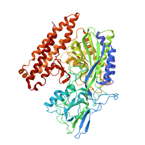
Human Glutamate Carboxypeptidase II Inhibition: Structures of Gcpii in Complex with Two Potent Inhibitors, Quisqualate and 2-Pmpa.

Mesters, J.R.Henning, K.Hilgenfeld, R.

(2007) Acta Crystallogr D Biol Crystallogr 63: 508

  • DOI: https://doi.org/10.1107/S090744490700902X
  • Primary Citation Related Structures: 
    2JBJ, 2JBK

  • PubMed Abstract: 

    Human glutamate carboxypeptidase II (GCPII) occurs in the central nervous system as well as in human prostate (where it is called prostate-specific membrane antigen; PSMA). Inhibitors of the enzyme have been shown to provide neuroprotection, but may also be useful for the detection, imaging and treatment of prostate cancer. Crystal structures were determined of the extracellular part of GCPII (amino-acid residues 44-750) in complex with two potent inhibitors, quisqualate and 2-PMPA (the strongest GCPII inhibitor to date), at resolutions of 3.0 and 2.2 A, respectively. In addition, models were constructed for binding of the inhibitors willardiine, homoibotenate, L-2-amino-4-phosphonobutanoic acid and L-serine-O-sulfate to the S1' site of the enzyme. The common denominator for high-affinity binding to the S1' site is the formation of two strong salt bridges.


  • Organizational Affiliation
    • Institute of Biochemistry, Center for Structural and Cell Biology In Medicine, University of Lübeck, Ratzeburger Allee 160, Lübeck 23538, Germany. mesters@biochem.uni-luebeck.de

Macromolecule Content 

  • Total Structure Weight: 82.73 kDa 
  • Atom Count: 5,914 
  • Modeled Residue Count: 687 
  • Deposited Residue Count: 707 
  • Unique protein chains: 1

Macromolecules

Find similar proteins by:|  3D Structure
Entity ID: 1
MoleculeChains  Sequence LengthOrganismDetailsImage
GLUTAMATE CARBOXYPEPTIDASE 2707Homo sapiensMutation(s): 0 
EC: 3.4.17.21
UniProt & NIH Common Fund Data Resources
Find proteins for Q04609 (Homo sapiens)
Explore Q04609 
Go to UniProtKB:  Q04609
PHAROS:  Q04609
GTEx:  ENSG00000086205 
Entity Groups
Sequence Clusters30% Identity50% Identity70% Identity90% Identity95% Identity100% Identity
UniProt GroupQ04609
Glycosylation
Glycosylation Sites: 7Go to GlyGen: Q04609-1
Sequence Annotations
Expand
Reference Sequence

Oligosaccharides

Help  
Entity ID: 2
MoleculeChains Length2D Diagram GlycosylationD Interactions
2-acetamido-2-deoxy-beta-D-glucopyranose-(1-4)-2-acetamido-2-deoxy-beta-D-glucopyranose
B, C, D
2N-Glycosylation
Glycosylation Resources
GlyTouCan: G42666HT
GlyCosmos: G42666HT
GlyGen: G42666HT
Entity ID: 3
MoleculeChains Length2D Diagram GlycosylationD Interactions
alpha-D-mannopyranose-(1-3)-beta-D-mannopyranose-(1-4)-2-acetamido-2-deoxy-beta-D-glucopyranose-(1-4)-2-acetamido-2-deoxy-beta-D-glucopyranose
E
4N-Glycosylation
Glycosylation Resources
GlyTouCan: G81315DD
GlyCosmos: G81315DD
GlyGen: G81315DD

Small Molecules

Ligands 5 Unique
IDChains Name / Formula / InChI Key2D Diagram3D Interactions
G88

Query on G88


Download:Ideal Coordinates CCD File
M [auth A](2S)-2-(PHOSPHONOMETHYL)PENTANEDIOIC ACID
C6 H11 O7 P
ISEYJGQFXSTPMQ-SCSAIBSYSA-N
NAG

Query on NAG


Download:Ideal Coordinates CCD File
J [auth A],
K [auth A],
L [auth A]
2-acetamido-2-deoxy-beta-D-glucopyranose
C8 H15 N O6
OVRNDRQMDRJTHS-FMDGEEDCSA-N
ZN

Query on ZN


Download:Ideal Coordinates CCD File
F [auth A],
G [auth A]
ZINC ION
Zn
PTFCDOFLOPIGGS-UHFFFAOYSA-N
CA

Query on CA


Download:Ideal Coordinates CCD File
H [auth A]CALCIUM ION
Ca
BHPQYMZQTOCNFJ-UHFFFAOYSA-N
CL

Query on CL


Download:Ideal Coordinates CCD File
I [auth A]CHLORIDE ION
Cl
VEXZGXHMUGYJMC-UHFFFAOYSA-M
Binding Affinity Annotations 
IDSourceBinding Affinity
G88 BindingDB:  2JBJ Ki: min: 0.2, max: 31 (nM) from 7 assay(s)
Kd: min: 0.4, max: 0.5 (nM) from 2 assay(s)
IC50: min: 0.3, max: 80 (nM) from 7 assay(s)
EC50: 0.62 (nM) from 1 assay(s)

Experimental Data & Validation

Experimental Data

  • Method: X-RAY DIFFRACTION
  • Resolution: 2.19 Å
  • R-Value Free:  0.238 (Depositor), 0.266 (DCC) 
  • R-Value Work:  0.192 (Depositor) 
  • R-Value Observed: 0.193 (Depositor) 
Space Group: I 2 2 2
Unit Cell:
Length ( Å )Angle ( ˚ )
a = 100.755α = 90
b = 130.982β = 90
c = 159.611γ = 90
Software Package:
Software NamePurpose
REFMACrefinement
HKLdata reduction
SCALEPACKdata scaling

Structure Validation

View Full Validation Report


Ligand Structure Quality Assessment 


Entry History 

Deposition Data

Revision History  (Full details and data files)

  • Version 1.0: 2006-12-18
    Type: Initial release
  • Version 1.1: 2011-05-08
    Changes: Version format compliance
  • Version 1.2: 2011-07-13
    Changes: Version format compliance
  • Version 2.0: 2020-07-29
    Type: Remediation
    Reason: Carbohydrate remediation
    Changes: Advisory, Atomic model, Data collection, Derived calculations, Other, Structure summary
  • Version 2.1: 2024-10-16
    Changes: Data collection, Database references, Structure summary