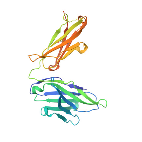
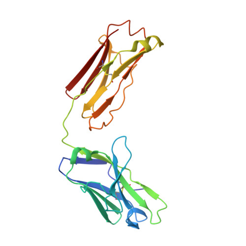
2JB5

Fab fragment in complex with small molecule hapten, crystal form-1


Experimental Data Snapshot

  • Method: X-RAY DIFFRACTION
  • Resolution: 2.80 Å
  • R-Value Free: 0.272 
  • R-Value Work: 0.211 

Starting Model: experimental
View more details

wwPDB Validation   3D Report Full Report


Ligand Structure Quality Assessment 


This is version 1.4 of the entry. See complete history


Literature

Fab Mor03268 Triggers Absorption Shift of a Diagnostic Dye Via Packaging in a Solvent-Shielded Fab Dimer Interface

Hillig, R.C.Urlinger, S.Fanghanel, J.Brocks, B.Haenel, C.Stark, Y.Sulzle, D.Svergun, D.I.Baesler, S.Malawski, G.Moosmayer, D.Menrad, A.Schirner, M.Licha, K.

(2008) J Mol Biol 377: 206

  • DOI: https://doi.org/10.1016/j.jmb.2007.12.071
  • Primary Citation of Related Structures:  
    2JB5, 2JB6

  • PubMed Abstract: 

    Molecular interactions between near-IR fluorescent probes and specific antibodies may be exploited to generate novel smart probes for diagnostic imaging. Using a new phage display technology, we developed such antibody Fab fragments with subnanomolar binding affinity for tetrasulfocyanine, a near-IR in vivo imaging agent. Unexpectedly, some Fabs induced redshifts of the dye absorption peak of up to 44 nm. This is the largest shift reported for a biological system so far. Crystal structure determination and absorption spectroscopy in the crystal in combination with microcalorimetry and small-angle X-ray scattering in solution revealed that the redshift is triggered by formation of a Fab dimer, with tetrasulfocyanine being buried in a fully closed protein cavity within the dimer interface. The derived principle of shifting the absorption peak of a symmetric dye via packaging within a Fab dimer interface may be transferred to other diagnostic fluorophores, opening the way towards smart imaging probes that change their wavelength upon interaction with an antibody.


  • Organizational Affiliation

    Bayer Schering Pharma AG, Global Drug Discovery, 13342 Berlin, Germany. roman.hillig@bayerhealthcare.com


Macromolecules
Find similar proteins by:  (by identity cutoff)  |  3D Structure
Entity ID: 1
MoleculeChains Sequence LengthOrganismDetailsImage
FAB FRAGMENT MOR03268 HEAVY CHAINA [auth H]244Homo sapiensMutation(s): 0 
Entity Groups  
Sequence Clusters30% Identity50% Identity70% Identity90% Identity95% Identity100% Identity
Sequence Annotations
Expand
  • Reference Sequence
Find similar proteins by:  (by identity cutoff)  |  3D Structure
Entity ID: 2
MoleculeChains Sequence LengthOrganismDetailsImage
FAB FRAGMENT MOR03268 LIGHT CHAINB [auth L]216Homo sapiensMutation(s): 0 
UniProt & NIH Common Fund Data Resources
Find proteins for P0DOY2 (Homo sapiens)
Explore P0DOY2 
Go to UniProtKB:  P0DOY2
GTEx:  ENSG00000211677 
Entity Groups  
Sequence Clusters30% Identity50% Identity70% Identity90% Identity95% Identity100% Identity
UniProt GroupP0DOY2
Sequence Annotations
Expand
  • Reference Sequence
Small Molecules
Ligands 1 Unique
IDChains Name / Formula / InChI Key2D Diagram3D Interactions
T5C
Query on T5C

Download Ideal Coordinates CCD File 
C [auth H]2-{(1E,3Z,5E,7E)-7-[3,3-DIMETHYL-5-SULFO-1-(2-SULFOETHYL)-1,3-DIHYDRO-2H-INDOL-2-YLIDENE]-4-METHYLHEPTA-1,3,5-TRIEN-1-YL}-3,3-DIMETHYL-5-SULFO-1-(2-SULFOETHYL)-3H-INDOLIUM
C32 H39 N2 O12 S4
WKWHOLCUESSZIQ-UHFFFAOYSA-O
Binding Affinity Annotations 
IDSourceBinding Affinity
TSC PDBBind:  2JB5 Kd: 0.6 (nM) from 1 assay(s)
Experimental Data & Validation

Experimental Data

  • Method: X-RAY DIFFRACTION
  • Resolution: 2.80 Å
  • R-Value Free: 0.272 
  • R-Value Work: 0.211 
  • Space Group: I 2 2 2
Unit Cell:
Length ( Å )Angle ( ˚ )
a = 71.949α = 90
b = 98.48β = 90
c = 153.782γ = 90
Software Package:
Software NamePurpose
CNXrefinement
HKL-2000data reduction
HKL-2000data scaling
MOLREPphasing

Structure Validation

View Full Validation Report


Ligand Structure Quality Assessment 


Entry History 

Deposition Data

Revision History  (Full details and data files)

  • Version 1.0: 2008-01-08
    Type: Initial release
  • Version 1.1: 2011-05-08
    Changes: Version format compliance
  • Version 1.2: 2011-07-13
    Changes: Version format compliance
  • Version 1.3: 2023-12-13
    Changes: Data collection, Database references, Other, Refinement description
  • Version 1.4: 2024-10-23
    Changes: Structure summary