X-ray structure of the T. aquaticus FtsY:GDP complex suggests functional roles for the C-terminal helix of the SRP GTPases.
Gawronski-Salerno, J., Coon 5th., J.S., Focia, P.J., Freymann, D.M.(2007) Proteins 66: 984-995
- PubMed: 17186523 Search on PubMedSearch on PubMed Central
- DOI: https://doi.org/10.1002/prot.21200
- Primary Citation Related Structures:
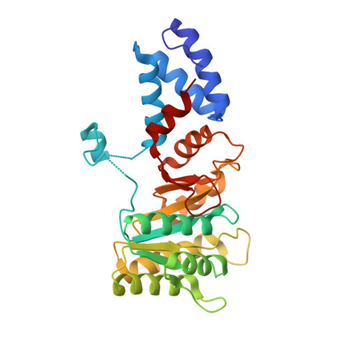
2IYL - PubMed Abstract:
FtsY and Ffh are structurally similar prokaryotic Signal Recognition Particle GTPases that play an essential role in the Signal Recognition Particle (SRP)-mediated cotranslational targeting of proteins to the membrane. The two GTPases assemble in a GTP-dependent manner to form a heterodimeric SRP targeting complex. We report here the 2.1 A X-ray structure of FtsY from T. aquaticus bound to GDP. The structure of the monomeric protein reveals, unexpectedly, canonical binding interactions for GDP. A comparison of the structures of the monomeric and complexed FtsY NG GTPase domain suggests that it undergoes a conformational change similar to that of Ffh NG during the assembly of the symmetric heterodimeric complex. However, in contrast to Ffh, in which the C-terminal helix shifts independently of the other subdomains, the C-terminal helix and N domain of T. aquaticus FtsY together behave as a rigid body during assembly, suggesting distinct mechanisms by which the interactions of the NG domain "module" are regulated in the context of the two SRP GTPases.
- Department of Molecular Pharmacology and Biological Chemistry, Northwestern University Medical School, Chicago, Illinois 60611, USA.
Organizational Affiliation: