2IYA | pdb_00002iya

The crystal structure of macrolide glycosyltransferases: A blueprint for antibiotic engineering


Experimental Data Snapshot

  • Method: X-RAY DIFFRACTION
  • Resolution: 1.70 Å
  • R-Value Free: 
    0.198 (Depositor), 0.198 (DCC) 
  • R-Value Work: 
    0.167 (Depositor), 0.167 (DCC) 
  • R-Value Observed: 
    0.168 (Depositor) 

wwPDB Validation 3D Report Full Report

Validation slider image for 2IYA

Ligand Structure Quality Assessment 


This is version 1.4 of the entry. See complete history

Literature

The Crystal Structure of Two Macrolide Glycosyltransferases Provides a Blueprint for Host Cell Antibiotic Immunity.

Bolam, D.N.Roberts, S.M.Proctor, M.R.Turkenburg, J.P.Dodson, E.J.Martinez-Fleites, C.Yang, M.Davis, B.G.Davies, G.J.Gilbert, H.J.

(2007) Proc Natl Acad Sci U S A 104: 5336

  • DOI: https://doi.org/10.1073/pnas.0607897104
  • Primary Citation Related Structures: 
    2IYA, 2IYF

  • PubMed Abstract: 

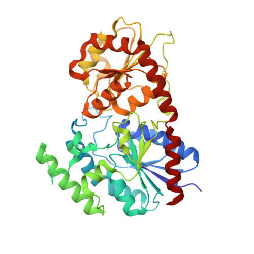
    Glycosylation of macrolide antibiotics confers host cell immunity from endogenous and exogenous agents. The Streptomyces antibioticus glycosyltransferases, OleI and OleD, glycosylate and inactivate oleandomycin and diverse macrolides including erythromycin, respectively. The structure of these enzyme-ligand complexes, in tandem with kinetic analysis of site-directed variants, provide insight into the interaction of macrolides with their synthetic apparatus. Erythromycin binds to OleD and the 23S RNA of its target ribosome in the same conformation and, although the antibiotic contains a large number of polar groups, its interaction with these macromolecules is primarily through hydrophobic contacts. Erythromycin and oleandomycin, when bound to OleD and OleI, respectively, adopt different conformations, reflecting a subtle effect on sugar positioning by virtue of a single change in the macrolide backbone. The data reported here provide structural insight into the mechanism of resistance to both endogenous and exogenous antibiotics, and will provide a platform for the future redesign of these catalysts for antibiotic remodelling.


  • Organizational Affiliation
    • Institute for Cell and Molecular Biosciences, The Medical School, University of Newcastle upon Tyne, Framlington Place, Newcastle upon Tyne NE2 4HH, UK.

Macromolecule Content 

  • Total Structure Weight: 93.61 kDa 
  • Atom Count: 6,690 
  • Modeled Residue Count: 776 
  • Deposited Residue Count: 848 
  • Unique protein chains: 1

Macromolecules

Find similar proteins by:|  3D Structure
Entity ID: 1
MoleculeChains  Sequence LengthOrganismDetailsImage
OLEANDOMYCIN GLYCOSYLTRANSFERASE
A, B
424Streptomyces antibioticusMutation(s): 0 
EC: 2.4.1
UniProt
Find proteins for Q3HTL7 (Streptomyces antibioticus)
Explore Q3HTL7 
Go to UniProtKB:  Q3HTL7
Entity Groups
Sequence Clusters30% Identity50% Identity70% Identity90% Identity95% Identity100% Identity
UniProt GroupQ3HTL7
Sequence Annotations
Expand
Reference Sequence

Small Molecules

Ligands 2 Unique
IDChains Name / Formula / InChI Key2D Diagram3D Interactions
ZIO

Query on ZIO


Download:Ideal Coordinates CCD File
D [auth A],
F [auth B]
(3S,5R,6S,7R,8R,11R,12S,13R,14S,15S)-6-HYDROXY-5,7,8,11,13,15-HEXAMETHYL-4,10-DIOXO-14-{[3,4,6-TRIDEOXY-3-(DIMETHYLAMINO)-BETA-D-XYLO-HEXOPYRANOSYL]OXY}-1,9-DIOXASPIRO[2.13]HEXADEC-12-YL 2,6-DIDEOXY-3-O-METHYL-ALPHA-L-ARABINO-HEXOPYRANOSIDE
C35 H61 N O12
RZPAKFUAFGMUPI-KGIGTXTPSA-N
UDP

Query on UDP


Download:Ideal Coordinates CCD File
C [auth A],
E [auth B]
URIDINE-5'-DIPHOSPHATE
C9 H14 N2 O12 P2
XCCTYIAWTASOJW-XVFCMESISA-N

Experimental Data & Validation

Experimental Data

  • Method: X-RAY DIFFRACTION
  • Resolution: 1.70 Å
  • R-Value Free:  0.198 (Depositor), 0.198 (DCC) 
  • R-Value Work:  0.167 (Depositor), 0.167 (DCC) 
  • R-Value Observed: 0.168 (Depositor) 
Space Group: P 1 21 1
Unit Cell:
Length ( Å )Angle ( ˚ )
a = 51.338α = 90
b = 103.471β = 94.82
c = 74.59γ = 90
Software Package:
Software NamePurpose
REFMACrefinement
MOSFLMdata reduction
SCALAdata scaling
SHELXphasing

Structure Validation

View Full Validation Report


Ligand Structure Quality Assessment 


Entry History 

Revision History  (Full details and data files)

  • Version 1.0: 2007-03-27
    Type: Initial release
  • Version 1.1: 2011-05-08
    Changes: Version format compliance
  • Version 1.2: 2011-07-13
    Changes: Version format compliance
  • Version 1.3: 2018-01-24
    Changes: Source and taxonomy
  • Version 1.4: 2024-05-08
    Changes: Data collection, Database references, Other