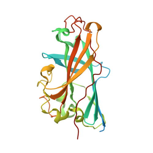
Structure of the C-terminal head domain of the fowl adenovirus type 1 long fiber.
Guardado-Calvo, P., Llamas-Saiz, A.L., Fox, G.C., Langlois, P., van Raaij, M.J.(2007) J Gen Virol 88: 2407-2416
- PubMed: 17698649
- DOI: https://doi.org/10.1099/vir.0.82845-0
- Primary Citation of Related Structures:
2IUM, 2IUN - PubMed Abstract:
Avian adenovirus CELO (chicken embryo lethal orphan virus, fowl adenovirus type 1) incorporates two different homotrimeric fiber proteins extending from the same penton base: a long fiber (designated fiber 1) and a short fiber (designated fiber 2). The short fibers extend straight outwards from the viral vertices, whilst the long fibers emerge at an angle. In contrast to the short fiber, which binds an unknown avian receptor and has been shown to be essential to the invasiveness of this virus, the long fiber appears to be unnecessary for infection in birds. Both fibers contain a short N-terminal virus-binding peptide, a slender shaft domain and a globular C-terminal head domain; the head domain, by analogy with human adenoviruses, is likely to be involved mainly in receptor binding. This study reports the high-resolution crystal structure of the head domain of the long fiber, solved using single isomorphous replacement (using anomalous signal) and refined against data at 1.6 A (0.16 nm) resolution. The C-terminal globular head domain had an anti-parallel beta-sandwich fold formed by two four-stranded beta-sheets with the same overall topology as human adenovirus fiber heads. The presence in the sequence of characteristic repeats N-terminal to the head domain suggests that the shaft domain contains a triple beta-spiral structure. Implications of the structure for the function and stability of the avian adenovirus long fiber protein are discussed; notably, the structure suggests a different mode of binding to the coxsackievirus and adenovirus receptor from that proposed for the human adenovirus fiber heads.
Organizational Affiliation:
Departamento de Bioquímica y Biología Molecular, Facultad de Farmacia, Universidad de Santiago de Compostela, Campus Sur, E-15782 Santiago de Compostela, Spain.


