2IUK | pdb_00002iuk

Crystal structure of Soybean Lipoxygenase-D


Experimental Data Snapshot

  • Method: X-RAY DIFFRACTION
  • Resolution: 2.40 Å
  • R-Value Work: 
    0.203 (Depositor) 
  • R-Value Observed: 
    0.203 (Depositor) 

wwPDB Validation 3D Report Full Report

Currently 2IUK does not have a validation slider image.


This is version 1.3 of the entry. See complete history

Literature

Crystal Structures of Vegetative Soybean Lipoxygenase Vlx-B and Vlx-D, and Comparisons with Seed Isoforms Lox-1 and Lox-3.

Youn, B.Sellhorn, G.E.Mirchel, R.J.Gaffney, B.J.Grimes, H.D.Kang, C.

(2006) Proteins 65: 1008

  • DOI: https://doi.org/10.1002/prot.21182
  • Primary Citation Related Structures: 
    2IUJ, 2IUK

  • PubMed Abstract: 

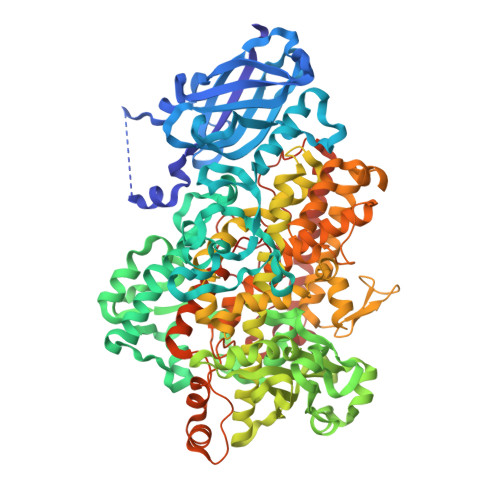
    The lipoxygenase family of lipid-peroxidizing, nonheme iron dioxygenases form products that are precursors for diverse physiological processes in both plants and animals. In soybean (Glycine max), five vegetative isoforms, VLX-A, VLX-B, VLX-C, VLX-D, VLX-E, and four seed isoforms LOX-1, LOX-2, LOX-3a, LOX-3b have been identified. In this study, we determined the crystal structures of the substrate-free forms of two major vegetative isoforms, with distinct enzymatic characteristics, VLX-B and VLX-D. Their structures are similar to the two seed isoforms, LOX-1 and LOX-3, having two domains with similar secondary structural elements: a beta-barrel N-terminal domain containing highly flexible loops and an alpha-helix-rich C-terminal catalytic domain. Detailed comparison of the structures of these two vegetative isoforms with the structures of LOX-1 and LOX-3 reveals important differences that help explain distinct aspects of the activity and positional specificity of these enzymes. In particular, the shape of the three branches of the internal subcavity, corresponding to substrate-binding and O(2) access, differs among the isoforms in a manner that reflects the differences in positional specificities.


  • Organizational Affiliation
    • School of Molecular Biosciences, Washington State University, Pullman, Washington 99164-4660, USA.

Macromolecule Content 

  • Total Structure Weight: 193.85 kDa 
  • Atom Count: 13,534 
  • Modeled Residue Count: 1,670 
  • Deposited Residue Count: 1,728 
  • Unique protein chains: 1

Macromolecules

Find similar proteins by:|  3D Structure
Entity ID: 1
MoleculeChains  Sequence LengthOrganismDetailsImage
SEED LIPOXYGENASE
A, B
864Glycine maxMutation(s): 0 
EC: 1.13.11.12 (PDB Primary Data), 1.13.11.58 (UniProt)
UniProt
Find proteins for P24095 (Glycine max)
Explore P24095 
Go to UniProtKB:  P24095
Entity Groups
Sequence Clusters30% Identity50% Identity70% Identity90% Identity95% Identity100% Identity
UniProt GroupP24095
Sequence Annotations
Expand
Reference Sequence

Small Molecules

Experimental Data & Validation

Experimental Data

  • Method: X-RAY DIFFRACTION
  • Resolution: 2.40 Å
  • R-Value Work:  0.203 (Depositor) 
  • R-Value Observed: 0.203 (Depositor) 
Space Group: P 1 21 1
Unit Cell:
Length ( Å )Angle ( ˚ )
a = 92.76α = 90
b = 115.1β = 112.34
c = 120.22γ = 90
Software Package:
Software NamePurpose
X-PLORrefinement
AMoREphasing

Structure Validation

View Full Validation Report

Currently 2IUK does not have a validation slider image.


Entry History 

Deposition Data

Revision History  (Full details and data files)

  • Version 1.0: 2006-10-11
    Type: Initial release
  • Version 1.1: 2011-12-28
    Changes: Database references, Derived calculations, Non-polymer description, Other, Version format compliance
  • Version 1.2: 2022-05-04
    Changes: Database references, Derived calculations
  • Version 1.3: 2024-06-19
    Changes: Data collection