2ITG | pdb_00002itg

CATALYTIC DOMAIN OF HIV-1 INTEGRASE: ORDERED ACTIVE SITE IN THE F185H CONSTRUCT


Experimental Data Snapshot

  • Method: X-RAY DIFFRACTION
  • Resolution: 2.60 Å
  • R-Value Work: 
    0.200 (Depositor) 

Starting Model: experimental
View more details

wwPDB Validation 3D Report Full Report

Validation slider image for 2ITG

This is version 1.5 of the entry. See complete history

Literature

The catalytic domain of human immunodeficiency virus integrase: ordered active site in the F185H mutant.

Bujacz, G.Alexandratos, J.Qing, Z.L.Clement-Mella, C.Wlodawer, A.

(1996) FEBS Lett 398: 175-178

  • DOI: https://doi.org/10.1016/s0014-5793(96)01236-7
  • Primary Citation Related Structures: 
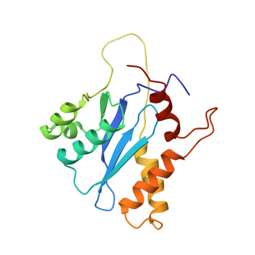
    2ITG

  • PubMed Abstract: 

    We solved the structure and traced the complete active site of the catalytic domain of the human immunodeficiency virus type 1 integrase (HIV-1 IN) with the F185H mutation. The only previously available crystal structure, the F185K mutant of this domain, lacks one of the catalytically important residues, E152, located in a stretch of 12 disordered residues [Dyda et al. (1994) Science 266, 1981-1986]. It is clear, however, that the active site of HIV-1 IN observed in either structure cannot correspond to that of the functional enzyme, since the cluster of three conserved carboxylic acids does not create a proper metal-binding site. The conformation of the loop was compared with two different conformations found in the catalytic domain of the related avian sarcoma virus integrase [Bujacz et al. (1995) J. Mol. Biol. 253, 333-346]. Flexibility of the active site region of integrases may be required in order for the enzyme to assume a functional conformation in the presence of substrate and/or cofactors.


  • Organizational Affiliation
    • Macromolecular Structure Laboratory, NCI-Frederick Cancer Research and Development Center, Frederick, MD 21702, USA.

Macromolecule Content 

  • Total Structure Weight: 17.92 kDa 
  • Atom Count: 1,291 
  • Modeled Residue Count: 160 
  • Deposited Residue Count: 163 
  • Unique protein chains: 1

Macromolecules

Find similar proteins by:|  3D Structure
Entity ID: 1
MoleculeChains  Sequence LengthOrganismDetailsImage
HUMAN IMMUNODEFICIENCY VIRUS-1 INTEGRASE163Human immunodeficiency virus 1Mutation(s): 1 
UniProt
Find proteins for P12497 (Human immunodeficiency virus type 1 group M subtype B (isolate NY5))
Explore P12497 
Go to UniProtKB:  P12497
Entity Groups
Sequence Clusters30% Identity50% Identity70% Identity90% Identity95% Identity100% Identity
UniProt GroupP12497
Sequence Annotations
Expand
Reference Sequence

Experimental Data & Validation

Experimental Data

  • Method: X-RAY DIFFRACTION
  • Resolution: 2.60 Å
  • R-Value Work:  0.200 (Depositor) 
Space Group: P 31 2 1
Unit Cell:
Length ( Å )Angle ( ˚ )
a = 72.4α = 90
b = 72.4β = 90
c = 65.56γ = 120
Software Package:
Software NamePurpose
PROLSQrefinement
DENZOdata reduction
SCALEPACKdata scaling

Structure Validation

View Full Validation Report


Entry History 

Deposition Data

Revision History  (Full details and data files)

  • Version 1.0: 1997-03-12
    Type: Initial release
  • Version 1.1: 2008-03-24
    Changes: Version format compliance
  • Version 1.2: 2011-07-13
    Changes: Derived calculations, Version format compliance
  • Version 1.3: 2021-11-03
    Changes: Database references, Other
  • Version 1.4: 2023-08-09
    Changes: Refinement description
  • Version 1.5: 2024-05-29
    Changes: Data collection